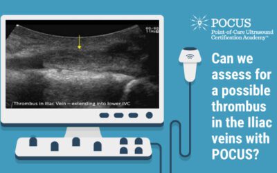
By Victor Rao MBBS, DMRD, RDMS - Abdomen, Ob/Gyn (APCA) Introduction Deep vein thrombosis (DVT) is a medical condition in which a thrombus develops in a deep vein, usually in the...
Inferior Vena Cava
POCUS in Acute Kidney Injuries
POCUS in Acute Kidney Injuries By Rolando Claure Del Granado Acute kidney injury (AKI) is a clinical syndrome caused by a multitude of hemodynamic, toxic, and structural insults...
Assessment for Central Venous Pressure
This infographic provides the steps required to perform a central venous pressure (CVP) assessment using point-of-care ultrasound (POCUS). Download the PDF: Discover more...
IVC: Upper Abdomen View
A 75-year-old female underwent abdominal ultrasound examination. The clinician observed that the IVC appeared suspicious. The following image and videos of the IVC in the upper...
POCUS: Differentiating the Aorta and IVC
Learn how to differentiate the aorta from the inferior vena cava during a point-of-can ultrasound scan. Want to grow in your application of POCUS in aortic aneurysm scenarios?...
IVC Anatomical Challenge
Case Study: A 28-year-old male athlete underwent an abdominal scan. The emergency medicine ultrasound fellow obtained the following sagittal view of the mid-IVC. What is the...
Inferior Vena Cava POCUS Assessment of Inferior Vena Cava (IVC)
Victor Rao MBBS, DMRD, RDMS (APCA) Anatomy The inferior vena cava (IVC) is a large, thin-walled, retroperitoneal blood vessel formed by the confluence of the left and right...
Inferior Vena Cava (IVC) Exam
Do you know how to detect the fluid status during a point-of-care-ultrasound examination of the inferior vena cava (IVC)? Normal IVC Note the difference in diameter of the IVC...


