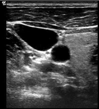
A 65-year-old male underwent a duplex ultrasound examination to assess the common carotid, external and internal carotid arteries. The internal jugular vein was also seen very clearly. No evidence of plaque or thrombus was seen in the vessels.
Which side carotid artery is displayed in the image below? The image orientation marker is indicated by the logo on the left upper corner of the image. The image is a transverse image of the common carotid artery. The image was obtained with the probe marker pointing towards the patient’s right side.
Which side carotid artery is displayed?
A. Left common carotid artery.
B. Right common carotid artery.
C. We cannot determine which side artery it is based on the image and the information provided.
The right common carotid artery is displayed.
Explanation
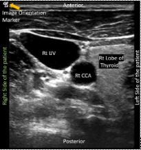
Figure 1. The transverse image of the right common carotid artery (RCCA) was obtained with the transducer orientation marker pointing towards the right side of the patient. The right lobe of the thyroid is seen just adjacent to the RCCA. See Figure 2 below showing the image obtained of the left common carotid artery (LCCA).
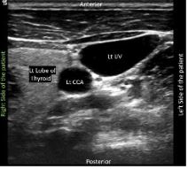
Figure 2. Left common carotid artery transverse view. An important fact to remember is the anatomical relationship of the thyroid lobe in relation to the common carotid artery. The lobe of the thyroid is more medial as compared to the LCCA in the image.
References
- https://www.isuog.org/static/uploaded/0e7c7719-b83d-4c36-b57f19ff777ab016.pdf
- https://wikem.org/wiki/Ultrasound:_Probe_orientation
- https://123sonography.com/blog/ultrasound-101-part-2-image-orientation
Access the full POCUS Learning Library for FREE!
Share a few details so we can tailor new content to your specialty and region.


