A 19-year-old female patient presented to the emergency room with a history of abdominal pain, nausea and vomiting for the past 2 days. The physician suspected the possibility of appendicitis and performed a POCUS examination. The following view was obtained.
What is the diagnosis?
A. Intussusception
B. Normal appendix diameter of less than 2.0 cm
C. Acute appendicitis as maximal diameter of the appendix at the widest point was more than 0.6 cm or 6 mm
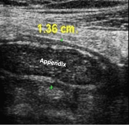
Image courtesy of UltrasoundCases.info, owned by SonoSkills.
Explanation
The mid longitudinal view of the appendix shows a dilated appendix with a diameter of 1.36 cm or 13.6 mm. The diameter is more than 6 mm, thus consistent with a diagnosis of acute appendicitis. Always measure from outer wall to outer wall as shown in the image.
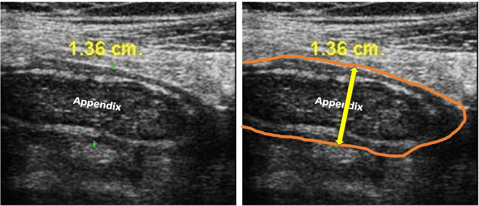
Figure. Appendix outer wall outlined in the image on the right.
References
- https://www.pocus101.com/abdominal-ultrasound-made-easy-step-by-step-guide/
- https://www.wjgnet.com/1949-8470/full/v3/i4/85.htm
- https://theultrasoundjournal.springeropen.com/articles/10.1186/2036-7902-5-S1-S2
- https://link.springer.com/article/10.1007/s00268-021-06077-5