A 71-year-old male presented with nausea, vomiting and right lower quadrant pain for 2 days. An abdominal POCUS exam was performed. The following images were obtained during the scan.
What is the diagnosis?
A. Intussusception
B. Acute appendicitis
C. Diverticulitis
Images courtesy of UltrasoundCases.info owned by SonoSkills
Explanation
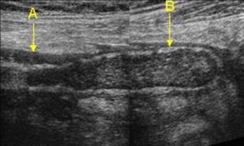
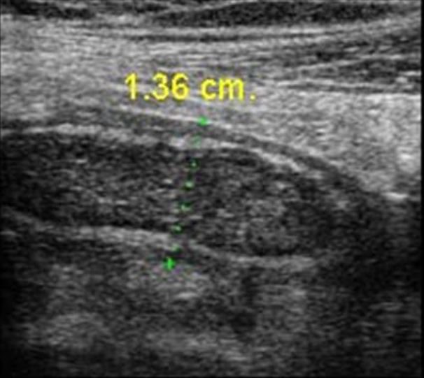
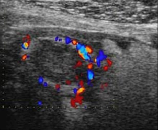
The B-mode images show a dilated non-compressible dilated appendix. Diameter of the appendix was 1.36 cm. Diameter of appendix more than 6 mm is considered a direct sign of acute appendicitis. No fecolith seen within the lumen of the appendix. No complex collection seen around the appendix. Small amount of fluid is seen around the appendix. Color Doppler image shows increased vascularity of the appendix (ring of fire) and target sign is positive in the transverse view.
References
Test your knowledge of Abdominal Trauma POCUS
with this knowledge check!