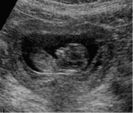
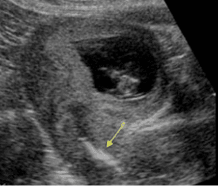
A 38-year-old female presented to the Emergency Department with history of slight vaginal bleeding. A pelvic ultrasound exam was performed. Single live intrauterine embryo was seen. Cardiac activity was observed. A linear echogenic structure was observed in the region of the cervical canal.
What is the arrow pointing to?
A. Blood clot
B. Air in the cervical canal
C. Intrauterine device (IUD)
Images courtesy of www.ultrasoundcases.info
Explanation
Intrauterine contraceptive devices are quite effective in preventing intrauterine pregnancy. Fewer than 1-2% of women with a copper or hormonal IUD get pregnant every year. Ultrasound is a great imaging modality to localize an IUD. In this example the pregnancy occurred because the IUD got displaced and slipped into the cervical canal. Seek proper medical care to manage this condition. Gentle removal of the IUD would be necessary. If the pregnancy occurred with an older IUD in the uterine cavity it may lead to a spontaneous abortion. Transvaginal ultrasound may be needed to visualize the IUD clearly.
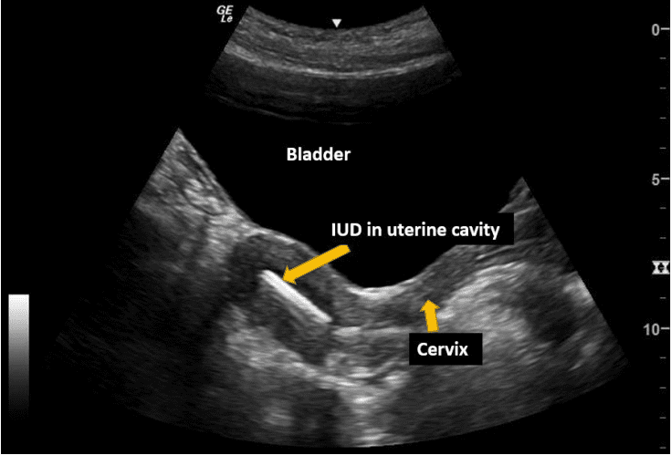
Figure – Normal location of an IUD in the uterine cavity in a non-pregnant patient.
References