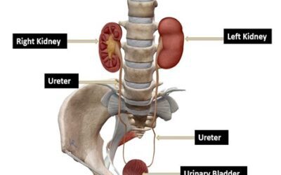
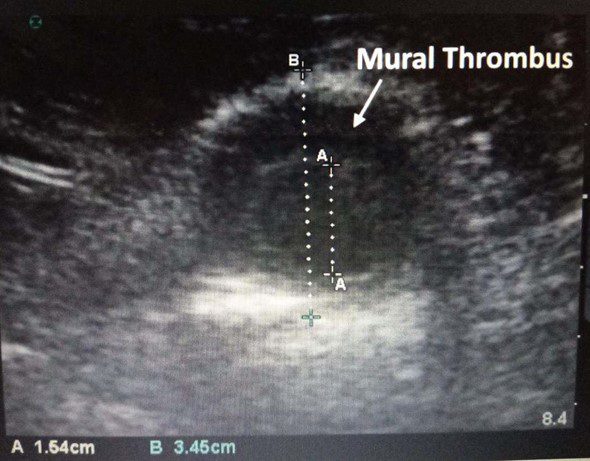
A 67–year–old male underwent an abdominal aorta aneurysm (AAA) screening ultrasound exam at the outpatient clinic. He had a history of smoking one pack of cigarettes for the past 40 years. He was also a known hypertensive. There was no known family history of AAA. The following image was obtained in the transverse view at the level of maximal dilatation of his lower abdominal aorta. Two measurements were performed.
What is the diameter of the aneurysm?
- 1.64 cm
- 3.45 cm
- None of the above
Explanation
The ultrasound image shows a transverse view of the lower abdominal aorta. The measurement should always be performed by placing the measurement calipers from outer wall to outer wall of the abdominal aorta. If a thrombus is present, include the thrombus also in the measurement. In this example, the AAA diameter is 3.45 cm.
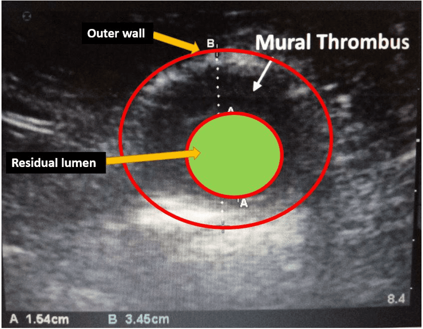
Figure 1. Transverse view of an abdominal aortic aneurysm at its widest level. Observe the caliper placement from outer wall to outer wall (A). You can measure the residual lumen, but the management decision is based on the outer wall to outer wall measurement. This can be a potential pitfall if you measure the residual lumen only resulting in a false negative.
References
- https://www.pocus101.com/aorta-ultrasound-made-easy-step-by-step-guide/
- doi.org/10.2310/6670.2008.00042
- doi.org/10.3390/biomedicines10010094
Test your POCUS AAA knowledge with this knowledge check!