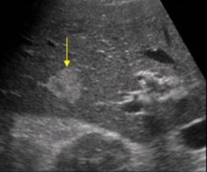
A 72-year-old male with a history of lung carcinoma underwent an ultrasound of the liver. The following image was obtained.
Images courtesy of UltrasoundCases.info owned by SonoSkills
What is the yellow arrow pointing to?
A. Liver metastasis
B. Fatty liver
C. Hemangioma
Test your knowledge of POCUS of Hepatobiliary and Spleen with this knowledge check!
Answer:
Liver hemangiomas typically are well defined hyperechoic lesions. They are seen quite commonly in the liver parenchyma and could be an incidental finding. Contrast enhanced ultrasound and biopsy of the lesion may be done if in doubt.