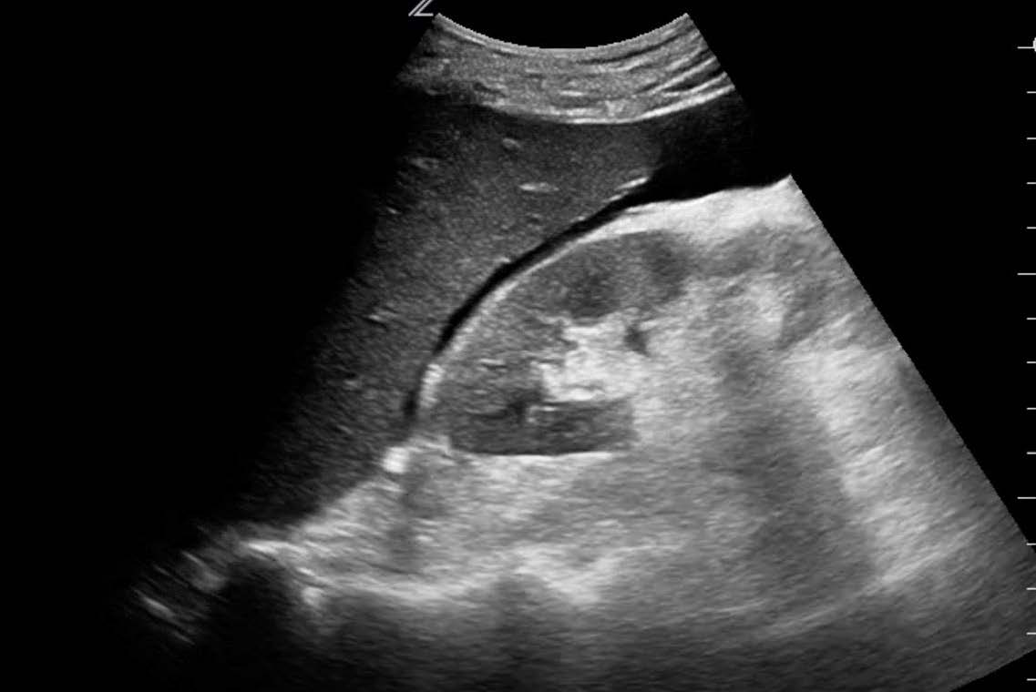
A 55-year-old female with a known history of breast carcinoma presented with right upper quadrant pain for 3 months. The physician decided to perform a right upper quadrant POCUS exam. The following images were taken during the right upper quadrant scan.
What is the most likely diagnosis?
A. Calcified echinococcus
B. Gallstones
C. Calcified liver metastases
Images courtesy of UltrasoundCases.info owned by SonoSkills
The most likely diagnosis is calcified liver metastases.
Explanation
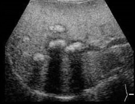
Ultrasound images show coarse echotexture of the liver with multiple hypoechoic lesions. The hypoechoic lesions have an echogenic area within them with a dense acoustic shadow. This is suggestive of calcified liver metastases. Further evaluation may be needed.
References
Test your knowledge of Hepatobiliary-Spleen POCUS
with this knowledge check!
Access the full POCUS Learning Library for FREE!
Share a few details so we can tailor new content to your specialty and region.


