A 45-year-old female patient presented with swelling in the left leg and thigh. The image below was obtained using a high frequency linear transducer. Power Doppler was also activated.
What is the most likely diagnosis?
A. Cellulitis
B. Edema
C. Abscess
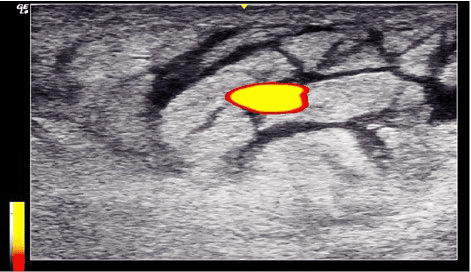
The most likely diagnosis is cellulitis.
Explanation
The image shows a cobble-stone appearance. Tissue architecture is not disrupted. No irregular collection with internal debris is seen. That rules out the possibility of an abscess in this view. On power Doppler, one prominent blood vessel is seen, but there is no evidence of profuse neovascularization.
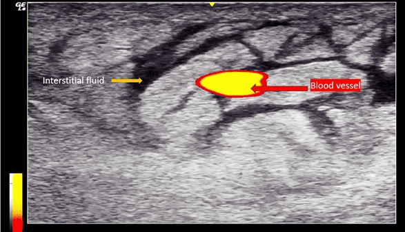
Ultrasound features of cellulitis are: increased thickness of the dermis, epidermis and subcutaneous tissue as compared to the normal side. Cellulitis and edema may sometimes look very similar. If the epidermis and dermis are abnormally hyperechoic then it is more suggestive of cellulitis. On the other hand, the epidermis and dermis does not appear very bright when simple edema is present.
Additionally, a negative compression test can be performed to look for large collections of fluctuant tissue and to rule out an abscess.
References
Loyer, E.M., DuBrow, R.A., David, C.L., Coan, J.D., Eftekhari. Imaging of superficial soft-tissue infections: sonographic findings in cases of cellulitis and abscess. Am J Roentgenol, 1996, 166(1): 149-152. https://doi.org/10.2214/ajr.166.1.8571865
Was this learning resource valuable?
Access the full POCUS Learning Library for FREE!
Share a few details so we can tailor new content to your specialty and region.