
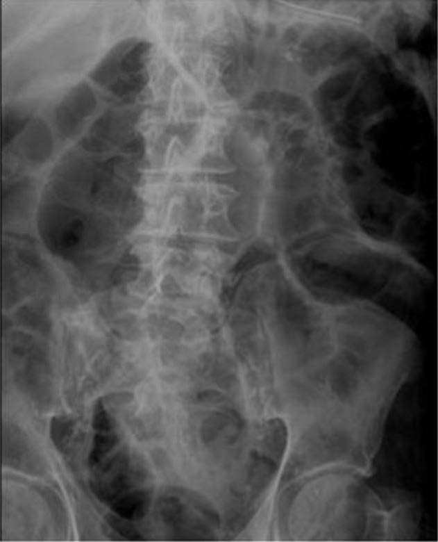
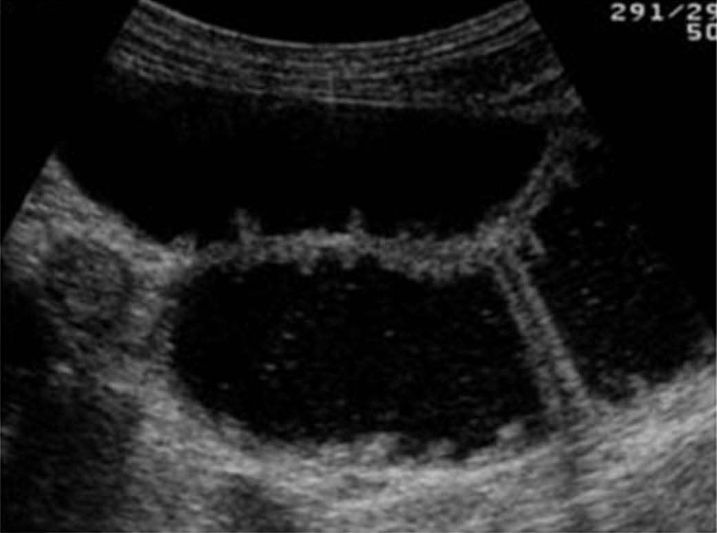
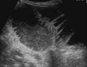
A 76-year-old male presented to the emergency department with a chief complaint of abdominal distension for 5 days. A plain X-ray abdomen was obtained. The physician also performed a POCUS examination of the abdomen.
What is the most likely diagnosis?
A. Pneumoperitoneum
B. Small bowel obstruction
C. Diverticulum
Images courtesy of UltrasoundCases.info owned by SonoSkills
Explanation
Ultrasound images show dilated fluid filled small bowel loops. Bowel wall appears to be thickened. Small bowel diameter was more than 2.5 cm. Bowel diameter measurement should be performed perpendicular to the bowel wall segment and should be measured from outer wall to outer wall. Some references mention small bowel diameter of more than 3.0 cm. More recent POCUS references consider a diameter of more than 2.5 cm as the criteria. Bowel content was seen moving to and fro (ineffective peristalsis). Correlate clinically and look for other findings which will confirm the diagnosis of SBO. X-ray abdomen shows predominantly central dilated small bowel with prominent valvulae conniventes in some bower segments.
References


