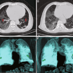
A 40-year-old male patient presented to the emergency department with a chief complaint of fatigue, mild fever, dyspnea, dry cough, chest, and muscle pain. He states that he returned from overseas a couple of days ago and is concerned he may have been exposed to the COVID-19 virus.
He has no significant past medical history and is not a known smoker. On physical exam, he had mild fever (99.6 degrees F) and an increased respiratory rate of 30 breaths per minute. A nasopharyngeal swab was collected and sent for PCR test for COVID-19.
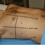
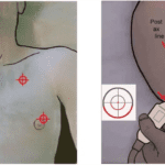
The following POCUS lung image was obtained in the intercostal space in zone 1 on his chest.
What does the image show?
A. Normal lung ultrasound
B. Multiple B-lines seen
C. Pneumothorax
D. A-lines seen
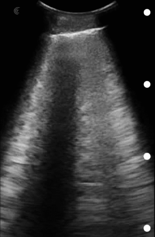
Multiple B-lines are seen – possibility of COVID-19 infection (to be confirmed on PCR).
Explanation
Presence of B-lines rules out the possibility of a pneumothorax in this intercostal space. There are 3 distinct B-lines seen through this intercostal space. More than 2 B-lines in a lung field imaged through an intercostal space are not considered to be normal. There may be occasional B-lines seen in older individuals in the basal region. No evidence of subpleural consolidation or air bronchograms were seen in this image.
References
1. Hasan AA, Makhlouf HA. B-lines: Transthoracic chest ultrasound signs useful in assessment of interstitial lung diseases. Ann Thorac Med. 2014;9(2):99-103. https://dx.doi.org/10.4103%2F1817-1737.128856</
Was this learning resource valuable?
Access the full POCUS Learning Library for FREE!
Share a few details so we can tailor new content to your specialty and region.