By Carlos Jimenez, DPT, RMSK, CPSS, PhD(c)
Lateral ankle sprains remain one of the most common injuries encountered in sports medicine, primary care, and emergency medicine settings. Approximately 85% of sprained ankles involve the lateral ligament complex, and most occur during sports or high-demand activities (1). Point-of-care ultrasound (POCUS) offers an immediate, ionizing radiation-free, and highly sensitive modality to evaluate these critical stabilizing structures in real time, complementing the physical examination and standard imaging.
This topic is particularly relevant for clinicians who perform musculoskeletal assessments including physical therapists, athletic trainers, sports physicians, emergency clinicians, and sonographers who rely on precise imaging to support clinical findings. In this post, we will provide an all-encompassing rundown of the lateral ankle ligament complex, including why ultrasound is a valuable tool for evaluating ATFL, CFL, and AITFL injuries and practical tips on how to perform a systematic scan and practical tips for clinicians who are new to musculoskeletal POCUS (2).
What Is the Lateral Ankle Ligament Complex and Why Does It Matter?
The lateral ankle ligament complex is composed of three primary stabilizing structures: the anterior talofibular ligament (ATFL), the calcaneofibular ligament (CFL), and the anterior inferior tibiofibular ligament (AITFL). These ligaments work together to limit excessive inversion, resist rotational forces, and maintain stability of the talocrural and subtalar joints during movement. Because of their anatomical orientation and the high mechanical demands placed on the ankle, they are among the most frequently injured ligaments in the body, particularly in sports and physically active populations (1).
Understanding this complex matters because up to 80–85% of ankle sprains involve the lateral ligaments, with the ATFL most commonly affected (1). Misdiagnosed or undertreated injuries can lead to chronic instability, impaired proprioception, recurrent sprains, and long-term degenerative changes (3).
For clinicians using POCUS, these ligaments are highly accessible and can be evaluated dynamically, making ultrasound a valuable tool for early diagnosis and targeted management (4). Whether ruling out a high-ankle sprain or confirming an isolated ATFL tear, accurate assessment of this ligament complex is essential for guiding rehabilitation, return-to-sport decisions, and patient education.
1. Anterior Talofibular Ligament (ATFL)
Purpose
Primary restraint to inversion and anterior translation of the talus in plantarflexion; most frequently injured ligament in lateral ankle sprains (1).
Probe Position
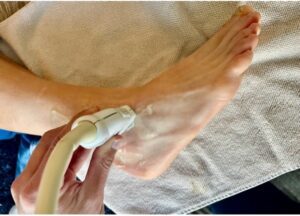
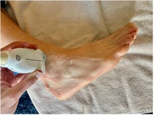
• Place the transducer in short axis to the distal fibula, angled slightly toward the talus.
• Maintain slight plantarflexion to relax overlying tissues.
Scanning Technique (2,5)
1. Identify the fibula on the lateral malleolus.
2. Slide anteriorly to visualize the talar neck.
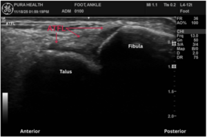
3. The ATFL appears as a thin, fibrillar, hyperechoic band spanning fibula to the talus.
4. Slowly sweep proximally and distally to assess the entire length.
Dynamic Assessment
• Apply a gentle anterior drawer or passive inversion.
• Assess ligament tension and symmetry with the contralateral side.
2. Calcaneofibular Ligament (CFL)
Purpose
Provides stability in dorsiflexion and resists inversion of the subtalar joint, second most injured ligament.
Probe Position
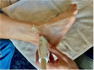
• Position the transducer inferior and slightly posterior to the fibular tip.
• Align the probe obliquely along the ligament toward the calcaneal tubercle.
Scanning Technique (2,5)
1. Locate the distal fibula and move inferiorly.
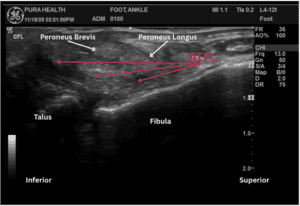
2. The peroneal tendons will be superficial; keep them centered but slightly anterior.
3. Deep to them, identify a thin, taut ligament running obliquely to the calcaneus.
4. Sweep along the ligament to assess continuity and thickness.
Dynamic Assessment
• Apply inversion stress in slight dorsiflexion.
• Observe for consistent tension throughout the ligament.
3. Anterior Inferior Tibiofibular Ligament (AITFL)
Purpose
Key stabilizer of the distal tibiofibular syndesmosis; commonly injured in high-ankle sprains or external rotation injuries.
Probe Position
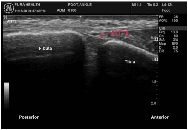
• Place the transducer horizontally across the anterior syndesmosis, just above the ankle joint.
• Align from anterolateral tibia to anteromedial fibula.
Scanning Technique
1. Identify the distal tibia medially.
2. Locate the distal fibula laterally.
3. The AITFL appears as a compact, fibrillar band connecting both bones.
4. Sweep along the ligament, ensuring you assess symmetry compared with the opposite side.
Dynamic Assessment
• Use external rotation stress or combined dorsiflexion and eversion.
• Look for syndesmotic widening, ligament elongation, or reproduction of the patient’s pain.
Analyzing Findings (Normal vs. Abnormal)
Once each ligament is visualized, interpretation centers on fiber continuity, echotexture, tension during dynamic maneuvers, and comparison with the opposite side. Normal ligaments appear thin, uniform, and tightly packed, with parallel echogenic fibers that remain taut during stress testing. Attachments to bone should be smooth and uninterrupted, with no surrounding fluid (2,3).
Abnormal findings vary by injury severity. Grade I sprains may show mild hypoechoic thickening without fiber disruption. Grade II injuries often display partial discontinuity, irregular fibers, or localized swelling. Complete tears demonstrate a clear gap between ligament ends, loss of tension with stress, and possible hypoechoic hematoma (3,4).
Dynamic testing enhances diagnostic confidence, for example, excessive talar translation for ATFL, reduced tension through the CFL, or widening of the syndesmosis for AITFL. Because ligament injuries can coexist with peroneal tendon pathology, joint effusion, or anterolateral impingement, a brief regional survey is recommended when abnormalities are present (4,6). Consistent side-to-side comparison helps distinguish true injury from normal anatomical variation.
Final Thoughts
Ultrasound offers clinicians a fast, dynamic, and highly accurate way to evaluate the lateral ankle ligament complex following acute or chronic injury. By combining a systematic scanning approach with thoughtful interpretation, POCUS users can differentiate between isolated ATFL sprains, combined ATFL-CFL injuries, and syndesmotic involvement each of which has different implications for recovery and return to activity (4,6). Whether used on the sideline, in the clinic, or as part of a comprehensive musculoskeletal examination, mastering the lateral ankle ultrasound assessment enhances diagnostic clarity and supports better patient care.
Call-to-Action
To continue developing your musculoskeletal ultrasound skills, explore additional educational resources, online learning modules, and certification pathways through the POCUS Certification Academy™. Strengthen your confidence, expand your clinical toolkit, and join a growing community of clinicians advancing point-of-care imaging.
References
1. Ferran NA, Maffulli N. Epidemiology of sprains of the lateral ankle ligament complex. Foot Ankle Clin. 2006. PubMed: 16971255
2. Jacobson JA. Fundamentals of Musculoskeletal Ultrasound. Elsevier. (Ankle ligament scanning protocols and sonoanatomy). Publisher page
3. Bianchi S, Martinoli C. Ultrasound of the Musculoskeletal System. Springer. Comprehensive ligament imaging and pathology). Book page
4. Halabchi F et al. Acute ankle sprain in athletes: Clinical aspects and algorithmic approach. World J Orthop. 2020. PubMed: 33362991
5. Precerutti M et al. Sonographic anatomy of the ankle. J Ultrasound. 2013. PubMed: 24883130
6. Sconfienza LM et al. Dynamic high-resolution US of ankle and midfoot ligaments. Radiographics. 2015. PubMed: 25590396
Was this learning resource valuable?
Access the full POCUS Learning Library for FREE!
Share a few details so we can tailor new content to your specialty and region.