By Victor Rao MBBS, DMRD, RDMS
Introduction
POCUS is an excellent tool for diagnosing and managing skin and soft tissue infections (SSTIs). It is primarily used to differentiate cellulitis from abscess, detect deeper tissue involvement, and guide ultrasound guided drainage if needed, and has higher diagnostic accuracy when the physical examination is equivocal.
A review of SSTIs diagnosed with ultrasound reported sensitivity in the 89–98% range and specificity 64–88%, with ultrasound consistently outperforming clinical examination alone in distinguishing an abscess from cellulitis. Ultrasound can also help detect serious life-threatening complications of SSTIs such as necrotizing fasciitis and gas gangrene which may have a higher incidence in patients with other comorbities.
Even though necrotizing fasciitis and gas gangrene are primarily clinical diagnoses, the physical examination alone is not sensitive enough to rule either one out. In this blog article we will mainly focus on cellulitis and abscess with some important ultrasound findings of gas gangrene which must never be missed because it can lead to serious complications and death.
Diagnostic Performance and Evidence
Systematic reviews and meta‑analyses of ED patients with SSTI show POCUS sensitivity around 90–97% and specificity about 80–83% for abscess vs cellulitis. In pediatric populations, pooled sensitivity is approximately 0.90 and specificity 0.80, outperforming physical examination alone (sensitivity 0.84, specificity 0.69).
POCUS frequently changes management, leading to correct management changes in about 10% of cases and improving detection of occult abscesses when the physical examination is ambiguous.
Characteristic Ultrasound Findings
On soft tissue ultrasound, abscesses classically show focal, relatively well‑defined hypoechoic to anechoic or heterogeneous fluid collection, often with internal echoes, debris, septations, and swirling material (snow‑globe or falling snow appearance) on compression. The debris is generally non-shadowing.
Posterior acoustic enhancement may be observed due to an increased through‑transmission through the liquified tissue. Surrounding cellulitis with cobble stoning of subcutaneous tissue with hypoechoic fluid between echogenic fat lobules, which may coexist with an abscess cavity.
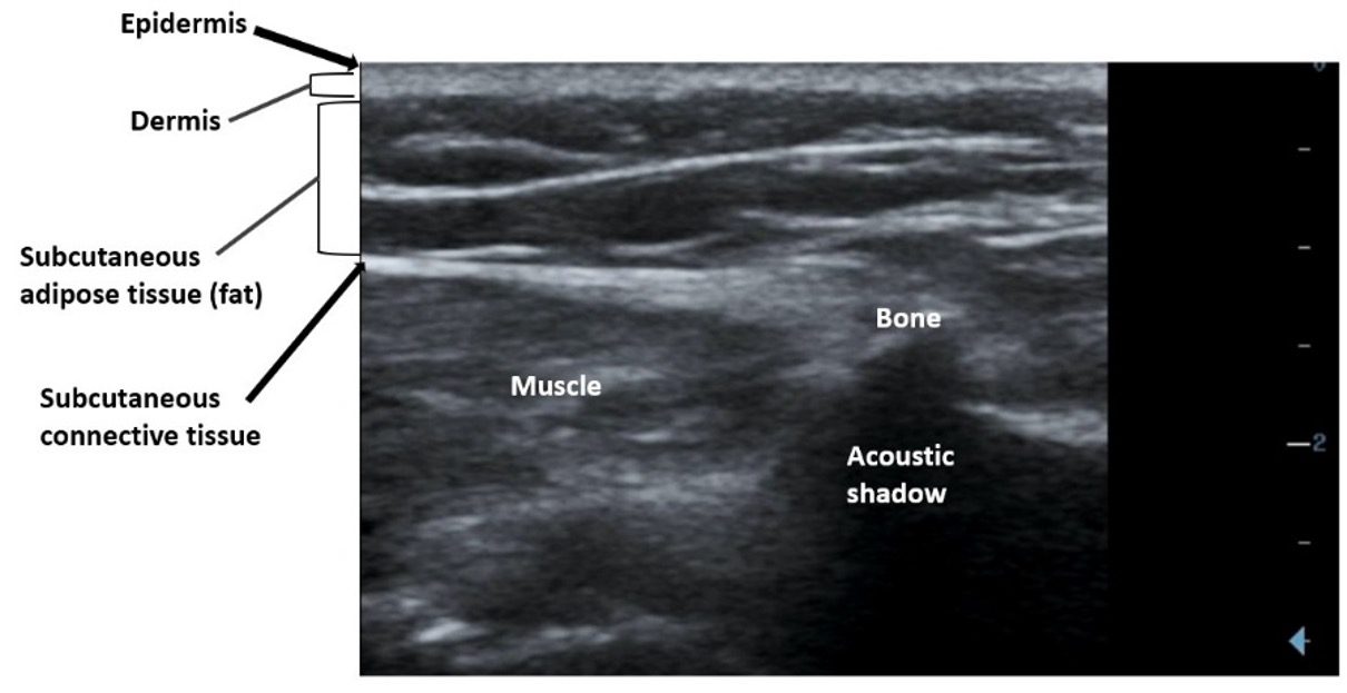
Figure 1. Layers of the skin and subcutaneous tissue on B-mode ultrasound.
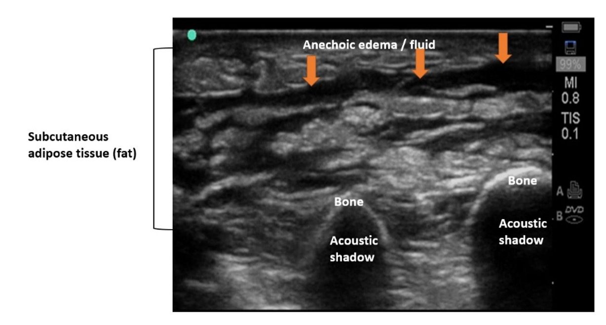
Figure 2. Cellulitis with hyperechoic fat lobules separated by hypoechoic fluid. Note that the architecture is still preserved. Compare with an abscess in figure 3.
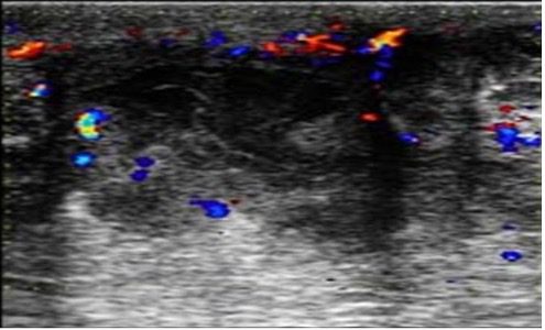
Figure 3. Abscess with an ill-defined heterogenous lesion with fluid pockets and debris. Color Doppler shows slightly enhanced color flow around the abscess.
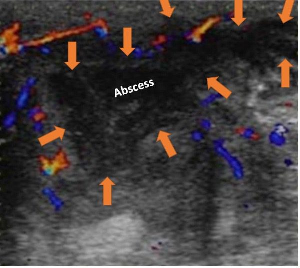
Figure 4. Abscess defined with orange arrows.
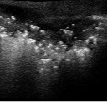
Figure 5. Necrotizing fasciitis and gas gangrene (Clostridial myonecrosis). The multiple bright lesions represent tiny pockets of air (which appear as punctate, hyperechoic inclusions) produced by gas-forming bacteria. Beyond the air bubbles there is a dirty shadow artifact blocking the view to deeper structures. Be aware that this is a life-threatening condition and must be managed immediately with surgical debridement and broad spectrum IV antibiotics. See figure 7 for comparison and labels.
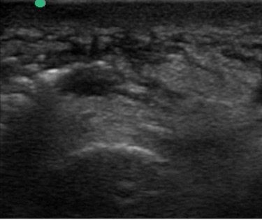
Figure 6. This is a tricky case. Hyperechoic subcutaneous fat lobules separated by hypoechoic fluid create a cobblestone appearance, consistent with cellulitis is seen. Observe the larger pocket of heterogeneous lesion below the cobblestone which is a small abscess. Two tiny air bubbles are also present which immediately change the diagnosis to gas gangrene in early stage and must not be missed as it can be fatal in not immediately managed appropriately. See figure 7 for labels and comparison.
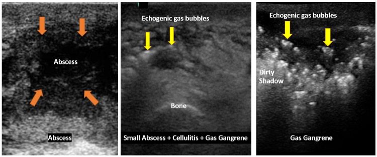
Figure 7. Image on the left shows an abscess in the subcutaneous fat region. Image in the middle shows two tiny air bubbles which should never be missed. The image on the right shows a more obvious and advanced stage of gas gangrene which cannot be missed.
Color Doppler will show peripheral hyperemia (Figures 3 and 4) but no flow within the cavity itself, helping to avoid confusion with lymph nodes or vascular structures. Gentle probe compression can demonstrate internal fluid movement or loculations, differentiating from solid masses.
Abscess Evaluation Technique and Tips
Use a high‑frequency linear transducer for superficial SSTI; curvilinear or phased‑array probes would be needed for deeper structures which are beyond the imaging rage of the high frequency linear transducer.
Systematically sweep in orthogonal planes (longitudinal and transverse) over and beyond the area of maximal tenderness, including contralateral normal side at the same anatomical level and plane for comparison. Optimize near‑field resolution for superficial lesions by adjusting the depth to include the entire cavity and underlying critical structures or using an acoustic standoff pad (ultrasound gel pad).
Always measure the abscess in three dimensions (length, width, depth) and record depth from skin surface to the superficial margin of the cavity. This is very valuable for follow up examinations to document if the abscess is decreasing in size as expected. Use color Doppler or power Doppler to detect significant vascular structures and map nearby vessels before incision or aspiration to avoid damaging the blood vessels.
Ultrasound Guided Management Principles
Incision and drainage vs conservative management
POCUS helps confirm or rule out an abscess when a physical examination is inconclusive, avoiding unnecessary incision and drainage (I&D) in cellulitis‑only cases and detecting occult collections that require drainage. Retrospective data suggests smaller, more superficial abscesses may be safely managed with antibiotics alone without immediate drainage.
One ED study found that patients with abscesses less than about 0.4–0.5 cm deep to the skin surface were more likely to receive and succeed with drainage, whereas deeper collections behaved differently. The authors proposed using size and depth thresholds to guide decisions.
Practical POCUS Thresholds
Very small abscess (<1 cm in diameter) and extremely superficial may resolve with antibiotics and close follow‑up if the patient is clinically well and has no fluctuance. Large abscess or those with significant loculations, systemic toxicity, or over high‑risk sites (face, hands, genitalia, near implants) generally warrant drainage and/or specialty consultation regardless of size.
Procedural guidance
Map out the extent and orientation of the abscess cavity, identifies safe skin entry points, and avoids blood vessels or critical adjacent structures. Ultrasound can guide needle aspiration or catheter placement, especially for deep soft tissue abscesses.
Ultrasound‑guided aspiration and irrigation of selected breast abscesses <3 cm has been shown to be preferable to surgical drainage in some series. Immediate post‑I&D ultrasound checks for residual loculations and guides further breakdown or additional incisions. Follow‑up ultrasound scans can document resolution of the abscess or increase in size.
| US Finding | Management | |
| Cellulitis | Cobblestone appearance only | Antibiotics only. Follow up US. No need for I&D |
| Large soft tissue abscess | Focal anechoic or heterogeneous collection with posterior acoustic enhancement, internal septations, debris | I&D + broad spectrum antibiotics. Wider incision and blunt exploration or catheter placement for drainage of pus |
| Small soft tissue superficial abscess | Small abscess with a diameter <1.0 cm | Conservative therapy, I&D, limited drainage may be considered if needed |
Table 1. Comparison of large soft tissue abscess, small soft tissue abscess and cellulitis ultrasound findings and management.
Final Thoughts
Point-of-care ultrasound (POCUS) is an effective clinical tool for diagnosing, distinguishing, and confirming soft tissue abscesses and cellulitis, thereby guiding appropriate management. This is a relatively easy skill to learn and should be used by all POCUS clinicians to manage the above-mentioned conditions.
Be aware of more serious conditions like necrotizing fasciitis and gas gangrene which require very aggressive treatment. Necrotizing fasciitis is a rapidly spreading, flesh-eating bacterial infection that fat and connective tissue under the skin. Gas gangrene invades and destroys muscle tissue and blood vessels and produces pockets of gas which will be seen on ultrasound as hyperechoic foci with reverberation artifacts (dirty acoustic shadows) See Figures 5, 6 and 7 and reference #5.
References
- Barbic D et al. Point‑of‑care ultrasound for diagnosis of abscess in skin and soft tissue infections: systematic review and meta‑ PMID: 27770490 DOI: 10.1111/acem.13049
- Gungor F et al. Role of point‑of‑care ultrasound in diagnosis of abscess in pediatric SSTI. PMID: 34379709 DOI: 10.11152/mu-3166
- Tsung JW et al. Abscess size and depth on ultrasound and association with treatment failure. J Emerg Med. 2020 Feb 26;21(2):336-342. doi: 10.5811/westjem.2019.12.41921. PMID: 32191191; PMCID: PMC7081847.
- O’Rourke K et al. Ultrasound for the Evaluation of Skin and Soft Tissue Infections. Mo Med. 2015 May-Jun;112(3):202-5. https://pubmed.ncbi.nlm.nih.gov/26168591/ PMID: 26168591; PMCID: PMC6170135.
- https://radiologykey.com/skin-and-soft-tissue/
Was this learning resource valuable?
Access the full POCUS Learning Library for FREE!
Share a few details so we can tailor new content to your specialty and region.