Submitted by Dr. Joana Ayuk, a third-year pediatric resident, Cameroon
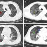
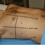
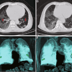
As a Pediatrics resident I decided to study POCUS because I realized how limited it was to rely solely on X-ray (chest X-ray particularly) for diagnosis, especially while managing unstable patients. So during one of my practical sessions, I performed a lung POCUS in a 14 year old adolescent who was treated for bilateral pneumonia, which was obvious on his initial chest X-ray.
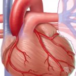
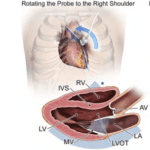
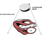
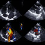
Despite proper management, his respiratory distress worsened especially when he laid down. So during my scan, lung POCUS was positive for bilateral pneumonia but surprisingly he had a moderate circumferential pericardial effusion. It explained his clinical state.
He was immediately transferred to the cardio thoracic service where a drainage was done. The fluid was purulent. Upon this new management, his respiratory distress subsided and with in 24 hours, his oxygen supply was reduced and a few days later, he was discharged.
POCUS helped save this boy’s life because it accurately saw where the anomaly was within minutes and completely changed the disease outcome.
Being in a low resource setting, I strongly believe POCUS is one of the indispensable tools we need to improve patient diagnosis and management in a setting where access to superior diagnostic tools aren’t always readily available.

Interview with Dr. Joana Ayuk
Tiffany Claiborne, Global POCUS Outreach Program Manager, Point-of-Care Ultrasound Certification Academy recently interviewed Dr. Joana Ayuk, diving further into the details of her shared POCUS story.
Q: To start, can you take us back to the 14-year-old boy you wrote about—the patient treated for bilateral pneumonia whose breathing worsened when he lay down? What was going through your mind at the bedside, and what led you to perform lung POCUS in that moment?
Sure. This was a 14 year old adolescent who was admitted for bilateral pneumonia and was treated with antibiotics. His chest X-ray was in accordance with initial diagnosis. 24 hours later, he was still very dyspneic and was put on oxygen support and that’s when I conducted lung POCUS because I thought it safe to exclude another underlying cause and bedside POCUS was the best tool I had at that instant.
Q: During that scan, you confirmed bilateral pneumonia, but you also discovered a circumferential pericardial effusion. How did that unexpected finding change your understanding of his condition and the decisions you made next?
This finding made us adjust our initial diagnosis as well as management. The patient’s life was in danger and we had to drain the pericardial fluid immediately. So we arranged for a quick transfer to the pediatric cardiothoracic service where drainage was done. The liquid was purulent. It was most likely that his pneumonia had also infected his pericardium because of their close contact. The patient was instantly relieved and discharged days later.
Q: After POCUS revealed the pericardial effusion and he was transferred for drainage, his respiratory distress improved and he was discharged days later. When you think about that sequence of events, how do you feel POCUS contributed to saving his life?
To be honest, this patient could have died if a lung POCUS was not conducted. The chest X-ray was not informative enough concerning the state of the pericardium – which is normal because a lung opacity is not different from the heart on X-ray. So having a tool that can instantly give us the state of the heart and the lungs is valuable to help guide our management. In our daily practice and especially in crashing patients, POCUS is a useful tool for quick decision making.
Q: You mentioned that relying only on chest X-ray can be limiting, especially with unstable patients. How has POCUS changed the way you approach diagnosis and management for children in your daily practice?
Ever since, I started studying POCUS, I’ve realized how important it is for clinicians to master it. Thanks to my friend and mentor Yannick Ndefo who inspired me through his determination at learning POCUS from scratch, and who graciously offered me his US apparatus for my practice of ultrasound, I’ve been able to safely put into practice all the teachings I’ve received. This has greatly contributed to the way I approach patients, especially the unstable ones. POCUS acts as a third eye that gives us a better insight into a patient’s state of health.
Q: In your experience, what are some of the ‘missed or hidden’ problems that POCUS has helped you uncover at the bedside that might not have been seen as quickly otherwise?
Concerning lung POCUS, an unexpected finding is a pericardial effusion in a patient with cough and fever. Lung POCUS can pick up very little effusions even before fluid starts to significantly build up. The same is true for small pleural effusions during pneumonia.
Q: You also shared that, in a low-resource setting, you believe POCUS is indispensable. What makes POCUS such a powerful tool where access to more advanced diagnostics isn’t always available, and what kinds of gaps does it help you bridge?
POCUS is readily available at the bedside and training is accessible with a rapid learning curve. If clinicians are well trained, they can better manage patients, especially in low income settings. POCUS does not replace other diagnostic tools, but it helps to guide who needs them urgently and who doesn’t. It gives us answers instantly on which we can rely to take the appropriate decisions. It’s time saving, with results instantly available.
Q: For clinicians and trainees who might be working in similar environments and wondering whether POCUS is worth the effort to learn, what message or encouragement would you share with them based on your experience?
If as a clinician you’ve been faced with a situation where you needed precise information on a crashing patient, and you were frustrated to be limited by lack of a readily diagnostic tool, you should quickly start your POCUS journey. I know it’s the case for many clinicians. Learning POCUS will give you answers instantly and guide your management of patients. My dream is to see fellow colleagues get involved and advocate for the widespread use of POCUS because it’s a tool that’s going to help improve our health system as a whole. While it takes determination, resources to learn are readily available so any health professional can easily access them, as well as participate in practical sessions organized by POCUS foundations and ambassadors.
Has POCUS impacted the care you provided to a patient? Has it altered the course of treatment or helped you to diagnose?
We want to hear your stories!
Was this learning resource valuable?
Access the full POCUS Learning Library for FREE!
Share a few details so we can tailor new content to your specialty and region.