By Victor Rao MBBS, DMRD, RDMS
Point-of-care ultrasound (POCUS) has become an essential tool in musculoskeletal medicine. It allows real-time, dynamic examinations without ionizing radiation, can guide management decisions, and procedures at the bedside. The MSK applications with the strongest evidence and the greatest impact on clinical decisions fall into the following categories:
Procedural Guidance
Joint aspiration and injections performed under ultrasound guidance markedly improve accuracy over landmark technique, especially in small or deep joints and fewer complications.
- Bursa and tendon sheath injections/aspirations
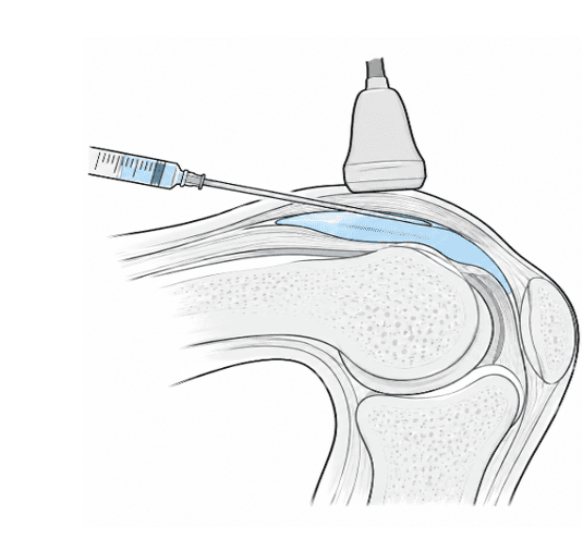
Figure 1. Diagrammatic representation of a knee joint aspiration/injection.
- Regional/peripheral nerve blocks
Ultrasound guided nerve blocks are now the standard of care for most nerve blocks in the emergency department and perioperative settings. See Figure 2.
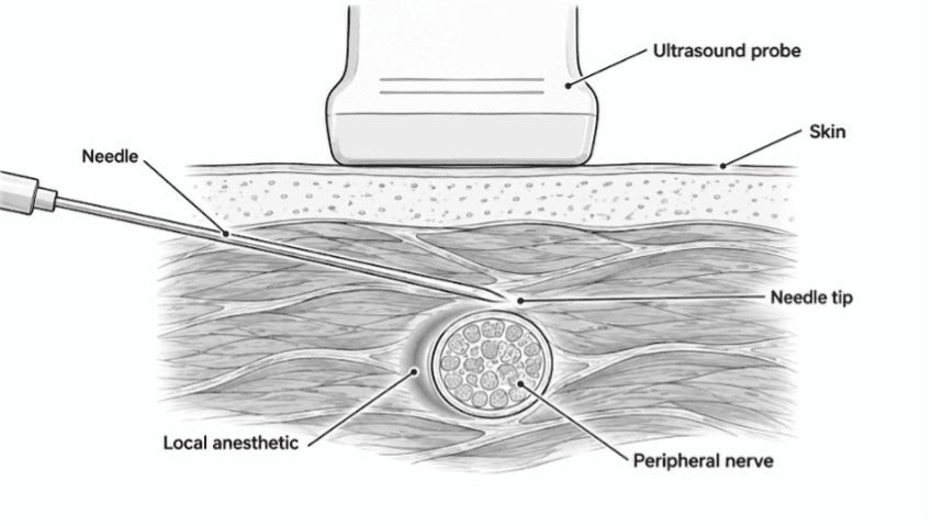
Figure 2. Diagrammatic representation of an ultrasound-guided nerve block. Direct sonographic visualization lets the operator deposit local anesthetic in the perineural space and avoids intraneural injection or needle trauma to the nerve.
Soft Tissue Infection Diagnosis
- Cellulitis versus abscess
Differentiating cellulitis from a drainable abscess (the classic “cobble stoning pattern” of cellulitis vs. an anechoic/heterogeneous fluid collection of an abscess). This directly changes management and guides incision and drainage.
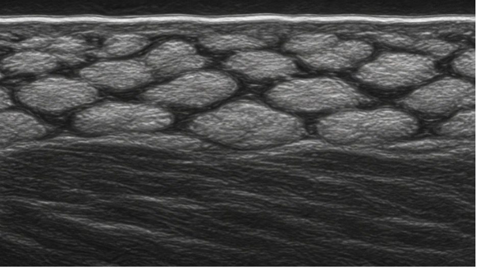
Figure 3. Diagrammatic representation of cobble stone pattern in cellulitis. Note that the tissue architecture is maintained. Cellulitis can be treated with broad-spectrum antibiotics and there is no indication to perform an incision and drainage.
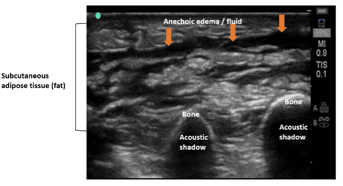
Figure 4. Cellulitis – note that the tissue architecture is preserved. See Figure 5 for comparison.
- Necrotizing fasciitis, gas gangrene and foreign body
Findings suggestive of necrotizing infection, gas gangrene or retained foreign material. Some findings could be very subtle in the early stages. Be vigilant. See figures 5 and 6.
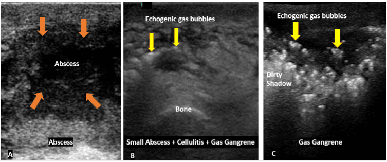
Figure 5. Image A shows a typical soft tissue abscess. Note the irregular ill-defined borders and a heterogeneous lesion. Image B shows a small abscess with two tiny hyperechoic air pockets. This should not be missed as this is consistent with gas gangrene. Figure C shows a more obvious case of gas gangrene. Note that both B and C are life-threatening conditions.
Foreign Body Detection
- Detecting foreign bodies embedded in soft tissue
Foreign bodies that are both radiopaque and radiolucent (such as wood splinter, glass, plastic, thorns) that plain X-ray films miss are easily detected by B-mode ultrasound.
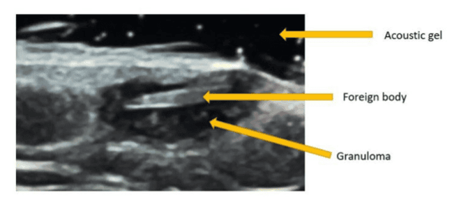
Figure 6. B-mode ultrasound image shows a linear hyperechoic foreign body with granulomatous tissue and fluid surrounding it.
Tendon and Muscle injury
- Full-thickness tendon tears
Achilles, quadriceps, patellar, distal biceps, and supraspinatus/rotator cuff. Dynamic scanning helps distinguish complete tears from partial tears.
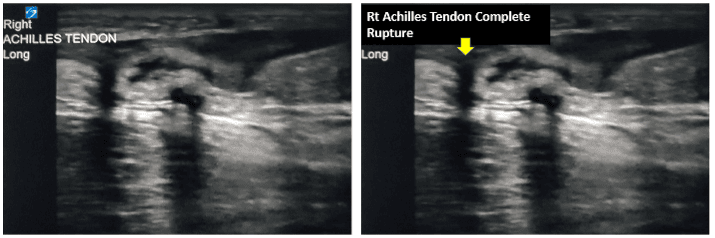
Figure 7. Complete Achilles tendon rupture. Ultrasound is a highly accurate diagnostic tool for evaluating Achilles tendon rupture with a sensitivity of up to 97% and specificity of 98%. Use a high frequency (10 to 15 MHz) linear transducer. Scan with the patient in the prone position with the feet hanging freely from the edge of the examination table. In this example there is complete disruption of the fibers of the Achilles tendon with an anechoic space seen between the torn ends (arrow). You may also gently do a passive dorsiflexion of the foot to make the lesion obvious if not clear. Always correlate clinically and compare with the normal side.
- Muscle tears, and hematomas
Ultrasound enables assessment of the normal architecture of muscles and tendon tears which could be partial or complete tears and dynamic ultrasound can help show the defect clearly. It can also identify hematomas which could vary in appearance with age.
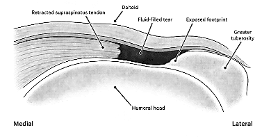
Figure 8. Diagrammatic representation of a complete supraspinatus tear.
Effusions and Joint Pathology
- Detecting and quantifying joint effusions
Detecting joint effusions and guiding aspiration. Pediatric hip effusion (transient synovitis vs. concern for septic arthritis) — a common and useful application.
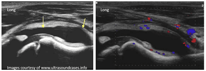
Figure 9. Coxitis Fugax (aseptic synovitis) A temporary, harmless inflammatory condition of the hip joint that frequently affects children aged 3 to 10 after a viral or upper respiratory infection. Patient presented with a limp. Ultrasound is very effective in diagnosing effusions around the joints.
Fracture Evaluation
- Detecting fractures
Detecting fractures where reducing radiation matters (such as patient’s in the pediatric age group) or where imaging is limited such as long bones, ribs, sternum and guiding fracture reduction in real time without the use of harmful ionizing radiation.
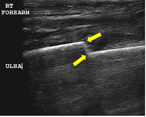
Figure 10. B-mode mid-longitudinal view of the ulna. Observe the focal discontinuity of the normally smooth, hyperechoic cortical line of the ulna in addition to a cortical step-off, and a small anechoic adjacent hematoma. Confirm and document the findings in both the mid-longitudinal and transverse planes. https://www.pocus.org/resources/cases/forearm-injury/
Nerve and Ligament Assessment
- Peripheral nerve entrapment
Peripheral nerve entrapment (e.g., median nerve in carpal tunnel). Ultrasound is an excellent imaging modality for diagnosing peripheral nerve entrapment. In carpal tunnel syndrome, it shows characteristic changes in the median nerve, such as enlargement at the carpal tunnel inlet, flattening within the tunnel, loss of the normal fascicular pattern, and reduced nerve mobility. See figure 11.
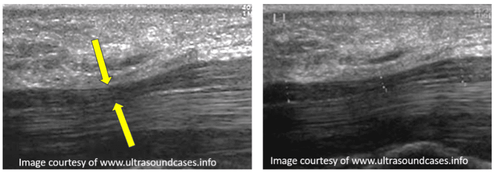
Figure 11. Narrowing/flattening of the median nerve in the carpal tunnel. You can measure as shown in the image on the right.
- Superficial ligament injuries
Superficial ligament injuries such as the thumb UCL and ankle ligaments can be effectively diagnosed with MSK POCUS.
Final Thoughts
MSK POCUS findings should support clinical judgement, not replace it. Use MSK POCUS judiciously for musculoskeletal assessment, procedural guidance and to speed up clinical diagnosis. My hope is that every clinician will make MSK POCUS part of routine practice to improve patient safety and deliver the best possible care. MSK POCUS is a vast subject. Each of the applications described above can be learned by any POCUS user, and proficiency can grow with continued training and experience.
*Disclaimer
AI (Perplexity Pro) and Claude were used to conduct extensive research on this topic and draft the outline and draw some of the diagrams. The author has reviewed, verified and edited the content for accuracy and relevance to POCUS primary care. Some views are the author’s own views but are based on consensus of the medical community.
Was this learning resource valuable?
Access the full POCUS Learning Library for FREE!
Share a few details so we can tailor new content to your specialty and region.