A 42-yr-old male presents to the emergency department with a chief complaint of lower abdominal pain and passage of blood in urine for 6 months. The physician performed a kidney and bladder ultrasound. The following images were obtained. Images were obtained in the transverse plane. Color Doppler was also performed.
What is the conclusion?
A. Bladder mass
B. Normal bladder ultrasound with ureteric jet
C. Bladder calculi with twinkle artifact and ureteric jet
Images courtesy of Dr. Kelechi Adioha
Bladder calculi with twinkle artifact and ureteric jet are present.
Explanation
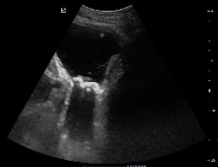
Ultrasound shows multiple hyperechoic calculi within the lumen of the bladder. The calculi are casting a dense acoustic shadow.
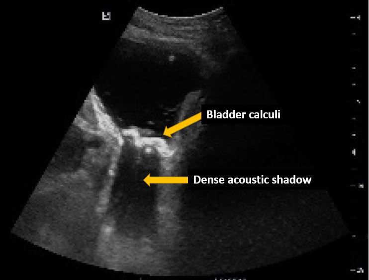
B-mode image of the bladder shows a distended bladder with hyperechoic calculi casting a dense acoustic shadow.
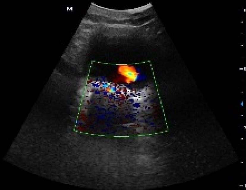
Color Doppler shows a small ureteric jet anteriorly and twinkle artifacts suggesting bladder calculi. Excessive color is also seen around the calculi due to high color gain. That can be corrected by reducing the color gain.
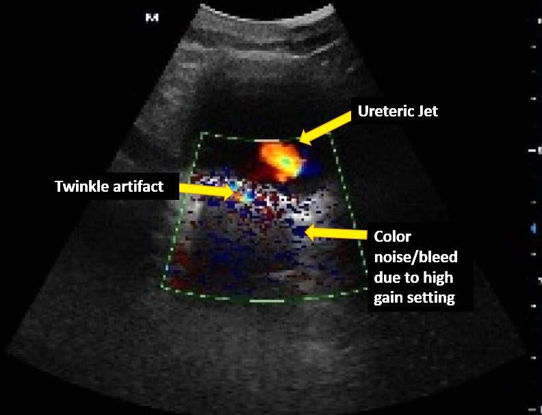
B-mode image with superimposed color Doppler shows a ureteric jet as the urine enters the bladder through the ureteric opening, twinkle artifact and some color noise/bleed due to excessive color gain. The noise can be reduced by lowering the color gain.
References
Test your knowledge of Renal/Genitourinary POCUS
with this knowledge check!


