
Case Study:
A 75-year-old male with PMH of HTN, DM Type II and ongoing tobacco use presented with complaint of worsening dyspnea, orthopnea, weight gain and lower extremity edema.
The patient also has a family history of ischemic heart disease, CVA. Temp – 98.4 degrees Fahrenheit, HR – 68 BPM, BP – 137/84 mm Hg, RR – 20 per minute. EKG shows – Q waves in inferior and lateral leads. No ST-T changes.
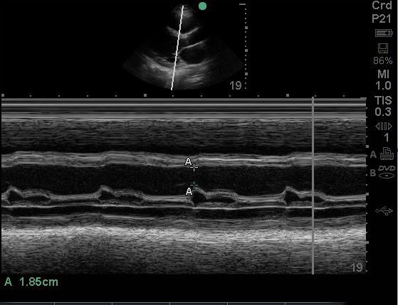
Figure showing the M-mode tracing in the PLAX view and the EPSS measurement.
Physical exam is remarkable for JVD, rales / crackles in lung bases, 1+ pitting edema up to the lower leg (bilateral). A POCUS cardiac exam was performed. An E-Point Septal Separation (EPSS) measurement was obtained. What is the approximate EF range based upon the EPSS measurement?
- The EF is more than 60%
- The EF is approximately 55%
- The EF is approximately 45%
- EF is less than 30%
Answer: 4: EF is less than 30%
Explanation:
The patient has severe LV systolic dysfunction. The approximate EF is less than 30%. Please see table below showing the approximate EF range based upon EPSS alone. Do keep in mind that there could be exceptions to this. One good example would be septal hypertrophy and mitral valve stenosis. Some experts are also able to estimate the EF using the eyeball method. But that judgement skill comes with significant experience.
| LVEF | EPSS Range (cm) |
| Normal >60% | <0.73 |
| Low normal (55-59%) | 0.74-0.89 |
| Mild systolic dysfunction (40-54%) | 0.9-1.39 |
| Moderate systolic dysfunction (30-39%) | 1.40-1.71 |
| Severe systolic dysfunction (<30%) | >1.71 |
A study using MRI data by Jay R Silverstein et al proposed that the mitral valve E point septal separation (EPSS) can be used to quantify LV ejection fraction on a continuous scale. They proposed the following formula to estimate EF on a continuous scale rather than just saying normal or reduced EF.
MRI LVEF = 75.5 – (2.5 x EPSS [millimeters])
Based upon the above equation, the EPSS is 29.25%. It agrees with the chart above as well. Note that this is still an approximate estimation of the EF. True EF can only be extrapolated from volumetric data.
Ready to begin your POCUS journey? Check out our many certificates and certifications here.


