
Case Study:
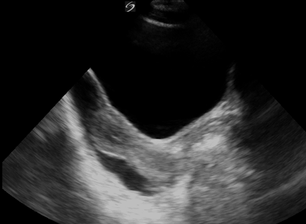
A 25-year-old female patient was brought to the emergency department (ED) with a history of being involved in a road traffic accident. A quick eFAST POCUS exam was performed in the ED. The following image was obtained while performing one of the views of the eFAST exam. The view obtained is a transabdominal view of the pelvic region in the mid-sagittal plane.
The image shows:
- Hydrosalpinx.
- Ovarian cyst.
- Normal physiological free fluid in the pelvic region.
- Anechoic free fluid present in the Pouch of Douglas and extending superior to the fundus of the uterus into the pelvic cavity.
Answer:
4. Anechoic free fluid present in the Pouch of Douglas and extending superior to the fundus of the uterus into the pelvic cavity.
Ready to get started on your POCUS journey? Check out our many certificates and certifications here.


