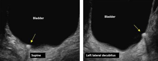
A 22-year-old male presented with colicky pain on the left side. He was diagnosed with left renal calculi in the past. The following ultrasound images were obtained during the bladder scan. The image on the left was obtained in the supine position and the image on the right was obtained after rolling the patient in the left lateral decubitus position.
What is the most likely diagnosis?
A. Calculus impacted at the right ureterovesical junction
B. Bladder wall calcification
C. Bladder calculus
Images courtesy of UltrasoundCases.info owned by SonoSkills
A bladder calculus is present.
Explanation
The image on the left shows a single round hyperechoic calculus/stone in the bladder. To confirm if the calculus is not impacted at the ureterovesical junction, roll the patient over to the left or the right lateral decubitus position. If the stone rolls to the dependent position, then it is within the lumen of the urinary bladder.
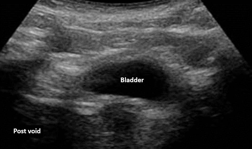
The post void bladder scan revealed no calculus in the bladder. The patient spontaneously passed the small bladder calculus during micturition.
References
Test your knowledge of Renal/Genitourinary POCUS
with this knowledge check!
Access the full POCUS Learning Library for FREE!
Share a few details so we can tailor new content to your specialty and region.


