
Case Study:
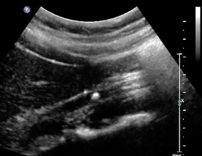
A 25-year-old male patient presented to the Emergency Department with a history of nausea, vomiting and pain in the right flank for the past 3 days. The patient took Tylenol tablets and had no relief. There was no significant past medical or family history. There was no history of chest pain or trauma. A quick POCUS abdominal scan was performed. The following image was obtained in the region of the right flank. What is the most likely diagnosis?
- Small bowel obstruction.
- Perforation of the bowel.
- Right ureteric calculus with right hydroureter.
- Appendicolith.
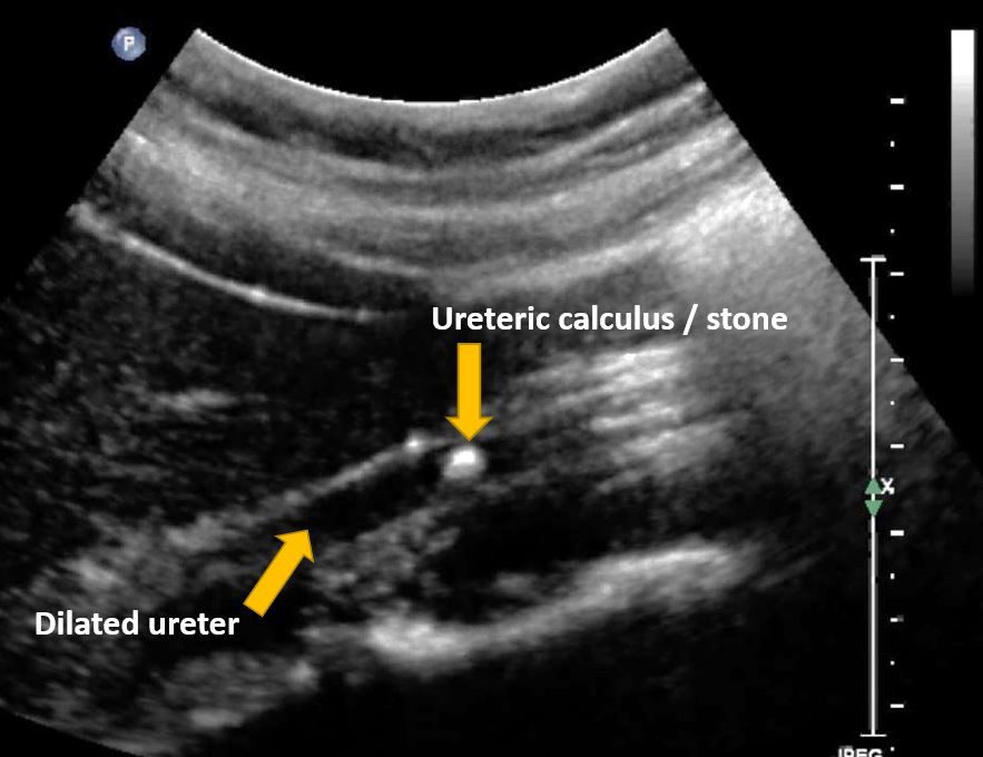
Answer: 3 Right ureteric calculus with right hydroureter
Explanation: The image obtained shows a long axis view through the right upper ureter. As seen in the image, the right ureter is dilated. A hyperechoic calculus with a faint posterior acoustic shadow is seen within the lumen of the right ureter. The ureter is generally hard to visualize on ultrasound when it is not dilated. However, when dilated due to an obstruction it is easy to visualize on ultrasound. You can also use color Doppler and overlay the color Doppler box in the region of the calculus to demonstrate the twinkle artifact. See labeled image below.
Ready to begin your POCUS journey? Check out our many Certificates and Certifications here.


