By Carissa Tomer, RDMS, RVT
Introduction
Diagnosing polycystic ovarian syndrome (PCOS) requires a combination of clinical and imaging findings in addition to laboratory test. If PCOS is suspected, a pelvic ultrasound is necessary. Point-of-care ultrasound (POCUS) can quickly image the ovaries and assess for signs of polycystic ovarian morphology.
With the increasing use of point-of-care ultrasound and the availability of quality in-office specialized ultrasound training, ovarian imaging has become a straightforward procedure that any gynecological provider can master with adequate training. This bedside examination enhances the practitioner’s understanding of the patient’s condition and helps rule out other underlying issues. This direct interaction fosters a stronger patient-provider connection and builds trust. Reports show patients feel more thoroughly examined when the provider performs a point-of-care ultrasound.
Furthermore, by incorporating POCUS into your practice as a gynecological provider, you can achieve timely diagnoses and implement treatment much faster. Consider this scenario: if a loved one needed an ultrasound and Provider A could perform it while others could not, we would all prefer Provider A. The time and money saved by the patient, coupled with the provider’s enhanced understanding of the patient’s condition and health, surpasses the outdated digital exam and referral process for external imaging. It is time to embrace the latest technology and move away from outdated practices.
There are both advantages and disadvantages to incorporating POCUS into your practice. The benefits include quicker diagnoses, more efficient treatment plans, cost savings for the patient, and less missed work time. As a provider, you will spend less time making follow-up calls and scheduling fewer follow-up visits. The only potential disadvantage is the small-time investment; if you cannot obtain the necessary bedside images, you have only invested a few minutes and can proceed with routine imaging as usual. However, this does not seem like a significant drawback to me.
How to Use POCUS for Evaluating the Ovaries
To evaluate the ovaries for polycystic ovarian morphology using POCUS, you will be looking for these findings:
- Follicle count: Are there more than 12 follicles?
- Ovarian size: Does the ovarian volume exceed 10 ml?
- Ovarian echotexture: Are the ovaries brighter than usual?
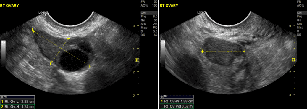
The image below shows a normal, healthy ovary with a single dominant follicle and a normal ovarian volume.
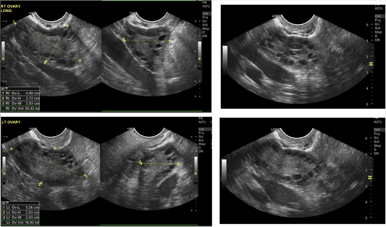
In the images below, a patient confirmed to have polycystic ovarian syndrome, you can appreciate numerous follicles referred to as a “string of pearls,” and the ovarian volume exceeds 10 ml. Compared to the normal ovary, the ovarian echotexture is brighter as well.
Conclusion
Even though diagnostic ultrasound imaging can diagnose suspicion for the possibility of polycystic ovarian syndrome; a comprehensive clinical assessment, menstrual history, evaluating hormone levels, and ultrasound findings, are all necessary to confirm the diagnosis.
Are you considering incorporating point-of-care ultrasound (POCUS) into your practice?
Ultrasound Energy is here to help. We specialize in assisting providers in seamlessly integrating POCUS into their practice. Contact us today to schedule your discovery session and explore how we can support you.
For those already performing POCUS and seeking to enhance their skills, we offer in-office education tailored to your needs.
Our mission is to raise awareness about the profound benefits of point-of-care ultrasound. We envision a healthcare system where all women receive a routine point-of-care pelvic ultrasound during every annual gynecologic visit, providing a comprehensive assessment that can significantly benefit women’s health.
Contact us today if you are ready to start offering POCUS in your practice; regardless of your specialty, we are here to help.
Contact Information
- Email: clientservices@ultrasoundenergy.com
- Phone: (941) 302-0663
- Website: Ultrasound Energy
Access the full POCUS Learning Library for FREE!
Share a few details so we can tailor new content to your specialty and region.


