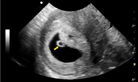
A 28-year-old female, G1P0 presented to the family medicine clinic for evaluation of her pregnancy. A transabdominal pelvic ultrasound was performed. The following is one of the images obtained.

What structure is the arrow pointing to?
A. Embryo
B. Yolk sac
C. Gestational sac
Explanation
The yolk sac is seen as an echogenic circular structure in the gestational sac. It can be visualized on ultrasound as early as 5.0 – 5.5 weeks gestation on transvaginal ultrasound. On a transabdominal scan it can be seen around 7.0 weeks gestation. It is the first definite sign of an intrauterine pregnancy. Embryo is not seen in this image. However, embryo was seen in the gestational sac while scanning in a different plane. An abnormal looking yolk sac or calcified yolk sac may suggest a bad outcome. Follow up ultrasound exams may be needed in those cases.
References
https://doi.org/10.1148/rg.2015150092
Want to test your Obstetrics POCUS knowledge further?
Try out our 7 question Obstetrics first trimester knowledge check!


