
A 35-year-old female presented to the Emergency Department with a chief complaint of pain, swelling and redness of the right thigh for the past 3 days. She gave a history of travel via a non-stop flight from Sydney to New York. She is currently also taking OCs. On inspection, it was observed that her right thigh was erythematous, and the girth was significantly increased as compared to the left side at the same level. The thigh was also warm to touch. A POCUS 2-zone compression test was performed bilaterally to assess for DVT. The two images below were obtained with and without compression at the level of the right saphenofemoral junction (SFJ).
What is the diagnosis?
A. Thrombus in the right common femoral vein
B. Thrombus in the right greater saphenous vein
C. Thrombus in the right common femoral vein and the right greater saphenous vein
Explanation
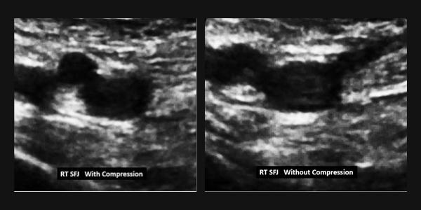
Consulting labeled Figure 1 below, the image on the left was obtained without compression at the right saphenofemoral junction. The common femoral artery (CFA), common femoral vein (CFV) and the greater saphenous vein (GSV) are seen. The image on the right was obtained with compression and shows that the greater saphenous vein is no longer visible because it is fully compressible.
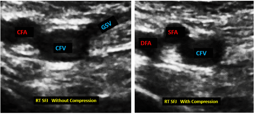
Figure 1. View of right saphenofemoral junction, without and with compression.
The common femoral vein (CFV) is not fully compressible and that suggests there is a thrombus within the CFV even though the thrombus is not visible on B-mode ultrasound. Remember that a thrombus can be anechoic, hypoechoic, or hyperechoic. Observe that the common femoral artery is not seen in the image on the right but instead we see two arteries (superficial femoral artery (SFA) and the deep femoral artery (DFA), also known as profunda femoris artery. The vessels sometimes tend to slip a little and we can instead see the divided CFA into the SFA and DFA (see colored image in Figure 2 below).
So, the correct answer is A. Thrombus in the right common femoral vein.
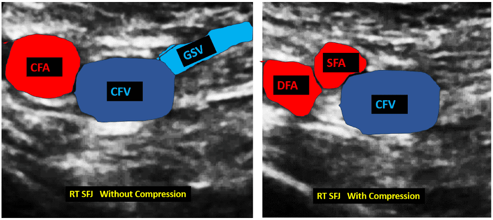
Figure 2. Observe that the greater saphenous vein (GSV) is no longer visible in the image on the right because it is fully compressible and thus has no thrombus in it. The common femoral vein (CFV) is not fully compressible in the image on the right which is consistent with a finding of a non-compressible CFV and thus a diagnosis of a thrombus in the CFV was made.
All images were obtained with the transducer orientation marker pointing towards the right side of the patient.
Want to test your DVT POCUS knowledge further?
Try out our 7 question DVT knowledge check!


