
The following image was obtained while scanning a patient in the late first trimester.
What is the diagnosis?
A. Missed abortion
B. Complete molar pregnancy
C. Partial molar pregnancy
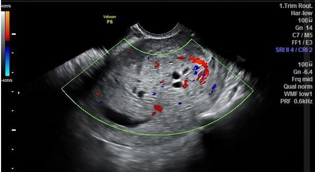
Image courtesy Radiopedia.org.
This is a partial molar pregnancy.
Explanation
The image shows an enlarged uterus with a small irregular gestational sac. Non-viable embryo was reported seen (not documented in the image). The placenta is thick and hyperechoic with multiple small anechoic spaces and increased blood flow on color Doppler. The ultrasound findings are consistent with partial molar pregnancy.
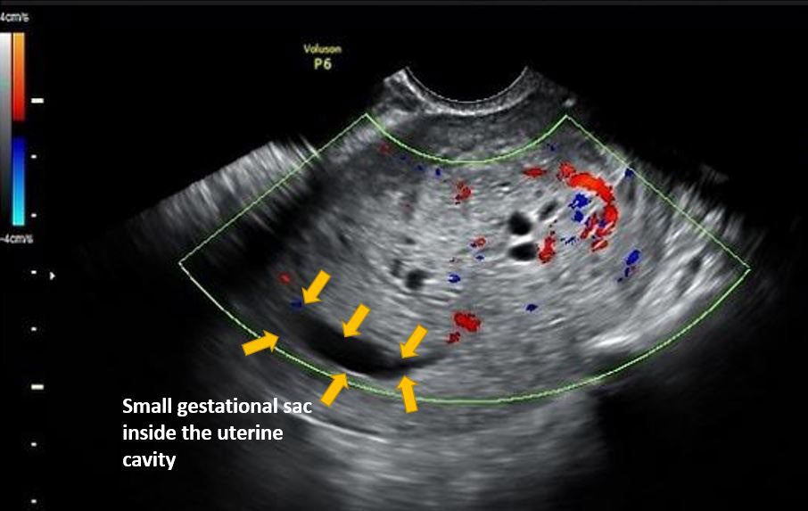
Figure – Partial molar pregnancy. Observe the irregular gestational sac (arrows). If a gestational sac (GS), embryo or embryo/fetus parts are seen along with the molar pregnancy cystic lesions then it is a partial molar pregnancy.
References
Test your knowledge of Obstetrics/First Trimester POCUS
with this 7 question quiz!


