Case Study:
A 52-year-old male patient underwent a POCUS exam to rule out gallstones. The gallbladder showed normal distension and the wall was not thickened. No pericholecystic fluid was seen. No evidence of gallstone or sludge was seen within the lumen of the gallbladder. An anechoic well defined circular cystic lesion was seen within the liver parenchyma. What is the most likely diagnosis?
What is the diagnosis?
A. Liver hemangioma
B. Anechoic free fluid in the Morison’s pouch
C. Dilated hepatic vein
D. Simple cyst of the liver
(Answer below)
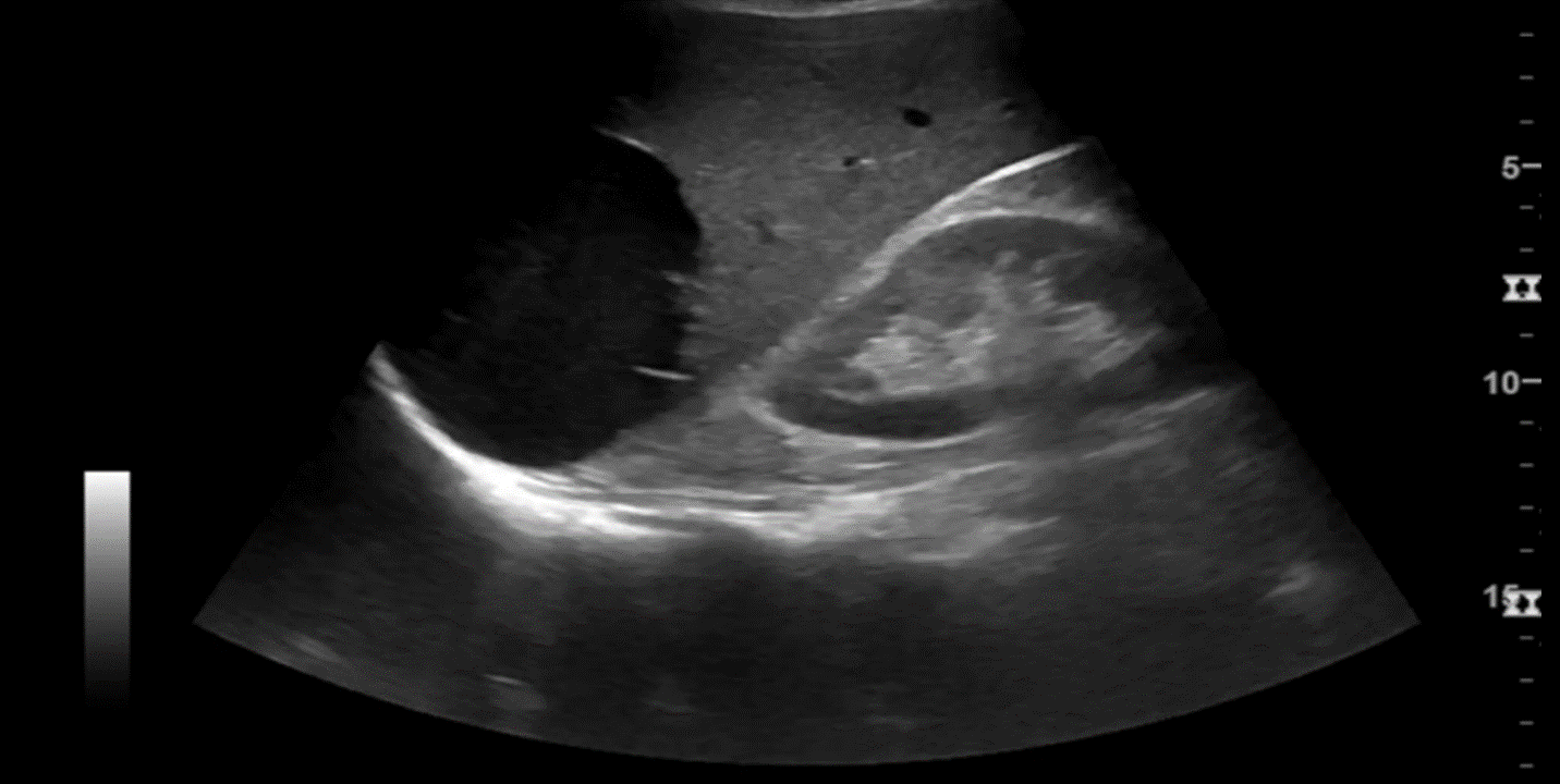
Answer: D, Simple cyst of the liver
Access the full POCUS Learning Library for FREE!
Share a few details so we can tailor new content to your specialty and region.


