
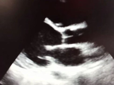
The following image was obtained while performing a limited cardiac POCUS examination. This is known as the parasternal long axis (PLAX) view of the heart.
What phase of the cardiac cycle is shown?
A. Systole
B. Diastole
C. Cannot be determined based on this view
The diastole phase of the cardiac cycle is shown.
Explanation
The transthoracic PLAX view of the heart shows the left atrium (LA), left ventricle (LV), right ventricular outflow tract (RVOT), anterior and posterior mitral valve (MV) leaflets, ascending aorta (AO) and the aortic valve (AV). Observe that the aortic valve is closed. The mitral valve is open. The blood should be rushing from the left atrium into the left ventricle (see red arrow). This is ventricular diastole.
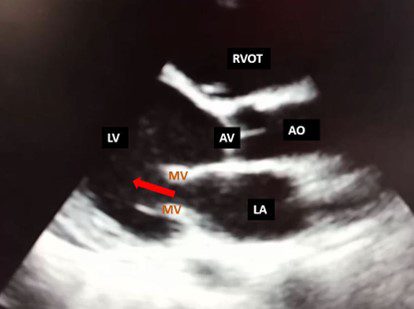
Figure 1. Observe direction of blood flow (red arrow) during diastole from the left atrium into the left ventricle.
References
- https://www.pocus101.com/cardiac-ultrasound-echocardiography-made-easy-step-by-step-guide/
- https://ecgwaves.com/topic/assessment-of-diastolic-function-by-echocardiography/
Test your knowledge further with our cardiac POCUS knowledge check. You’ll receive a score plus feedback on your answers.


