A 50-year-old female presented to the hospital with a history of chronic abdominal pain for 6 months.
She is a known alcoholic and had been diagnosed with acute pancreatitis in the past. The physician on duty performed an abdominal ultrasound. Below is one of the images of the pancreas.
What is the most likely diagnosis?
A. Pancreatic cancer
B. Chronic pancreatitis
C. Acute on chronic pancreatitis
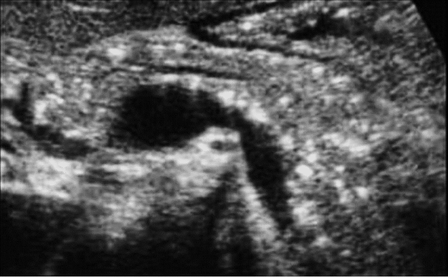
Image courtesy of UltrasoundCases.info, owned by SonoSkills.
Pancreatitis is the most likely diagnosis.
Explanation
The pancreas appears hyperechoic. The pancreatic duct is slightly dilated. Multiple hyperechoic calcifications are seen within the pancreatic tissue. The ultrasound findings are suggestive of chronic pancreatitis. No hypoechoic areas were seen in the pancreas.
Let us observe the image of a normal pancreas.
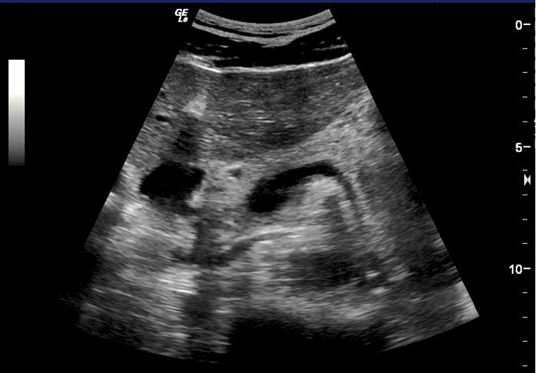
Figure 1. Normal pancreas (adult male).
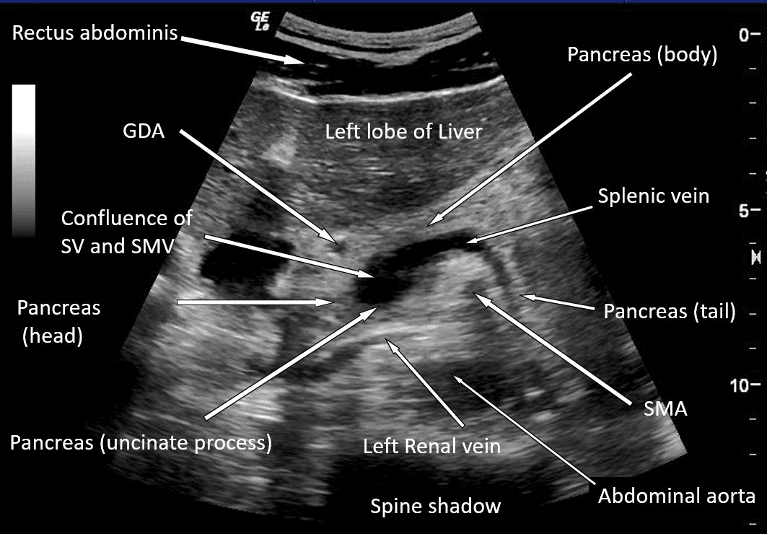
Figure 2. Labeled normal pancreas and important structures in its vicinity.
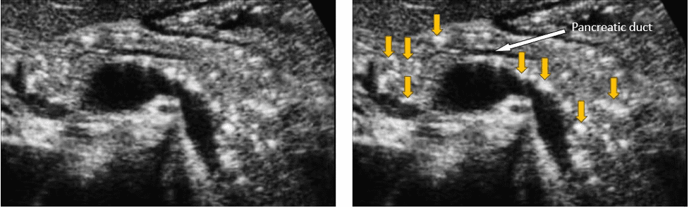
Figure 3. Slightly dilated pancreatic duct and multiple calcifications in the pancreas (yellow arrows). The pancreas appears hyperechoic as compared to the normal adult pancreas.
References
Access the full POCUS Learning Library for FREE!
Share a few details so we can tailor new content to your specialty and region.


