
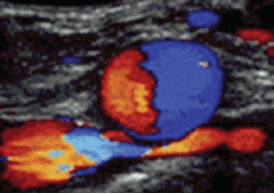
A 25-year-old female presented to the clinic with the chief complaint of pain and swelling in the left leg and thigh. She had no history of trauma. The following image was obtained of the right common iliac artery.
What is the likely diagnosis?
A. Normal anatomy
B. DVT in the left common iliac vein
C. May-Thurner syndrome
Explanation
May-Thurner syndrome is an anatomical variant that may predispose some individuals to develop a DVT.
This Color Doppler image shows the right common iliac artery (RCIA) crossing anterior to the left common iliac vein (LCIV) and compressing it:
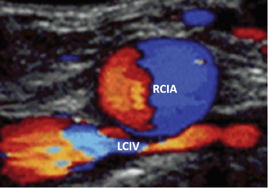
This results in partial venous flow obstruction (stasis) and creates an ideal site for thrombus formation. Individuals with this condition could be asymptomatic or may present with pelvic pain, left lower extremity swelling and varicose veins in the left leg. In post thrombotic patients these individuals may present with chronic leg ulcers, varicose veins, and pigmentation in the left lower extremity.
Initially, in stage 1, there is common iliac vein compression without any structural changes. In stage 2, there is venous spur formation and patients may present with edema and or DVT in the left lower extremity. Stage 3 presents as a DVT in the left lower extremity along with edema and some late changes such as varicose veins. Patients with a DVT may benefit from a stent placement in the region of stenosis. However, the DVT may recur on the left lower extremity.
Interested in learning more about May-Thurner syndrome?
Read the May-Thurner syndrome blog.
Want to test your DVT POCUS knowledge further?
Try out our 7 question DVT knowledge check!


