Case Study:
A 45-year-old female patient presented with a history of tiredness and dyspnea on exertion for past 1 year. On physical exam the patient was short of breath and had pitting edema in both lower extremities around her ankles. On auscultation a pan systolic murmur was heard which was best heard in the apical region. A POCUS cardiac exam was performed by the physician and the following was an apical 4 chamber view obtained with the color Doppler box overlay in the region of the mitral valve and the left atrium.
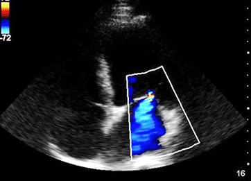
Figure showing color Doppler placed over the LA, MV and small segment of the LV.
What is the most likely diagnosis?
A. Normal color Doppler flow of blood from the left atrium into the left ventricle during ventricular diastole
B. The blue color indicates deoxygenated blood
C. The blue color indicates reverse flow from the left ventricle into the left atrium and is consistent with mitral valve regurgitation
D. The blue color suggests normal flow into the LA during atrial filling
Interested in proving your expertise in Cardiac POCUS? Check out our Cardiac POCUS Certificate here.
Answer: C
Explanation: When analyzing color Doppler flow display, always look at the legend first. In the above image it is located on the left upper corner of the image. The legend by default is red above and blue below the baseline and indicating flow towards and away from the transducer, respectively. It does not indicate arterial or venous flow. The blood flow into the left atrium (LA) from the left ventricle (LV) through the mitral valve (MV) is not normal. However, keep in mind that the MV may show small amount of reverse flow from the LV into the LA even in normal individuals. But, in the example above it appears to be significant.
Access the full POCUS Learning Library for FREE!
Share a few details so we can tailor new content to your specialty and region.


