A 35-year-old male presented with dyspnea and chest pain for 3 months.
After performing a physical examination, the physician decided to perform a POCUS lung ultrasound. The following image was obtained in the region of the right lower chest.
What structure is the arrow pointing to in the image?
A. Diaphragm
B. Lung
C. Heart
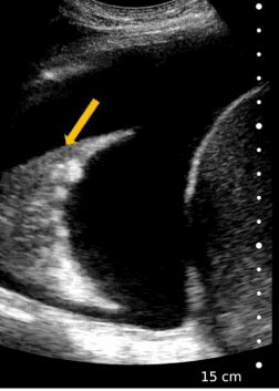
Image courtesy of Dr.Juraj Smaha
The arrow is pointing to the lung.
Explanation
The B-mode image shows a large anechoic pleural effusion. The spine sign is also positive as the large fluid collection collapsed the lung and pushed it superiorly. The diaphragm is also seen as a curved echogenic line. Jellyfish sign is also positive. There is compressive atelectasis of the lung parenchyma, which was seen moving in a large pleural effusion, which is indicative of a low viscosity of pleural fluid.
No debris or septation/adhesions seen within the fluid.
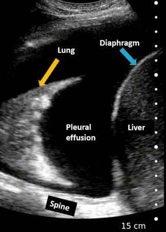
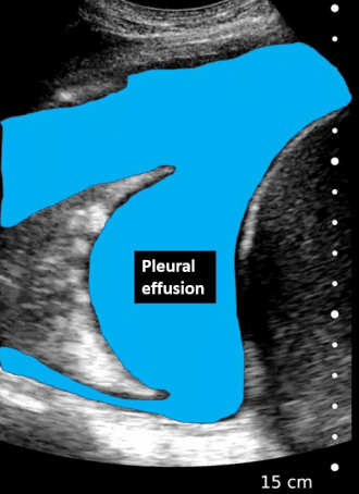
Figure – Observe the collapsed lung with small echogenic air bronchograms (white areas near the base of the lung). Note that the spine is visible, which is normally not visible in this view because of the aerated lung anterior to it. The large pleural effusion created a perfect acoustic window to allow visualization of the spine.
References
Test your POCUS lung knowledge with this knowledge check!
Access the full POCUS Learning Library for FREE!
Share a few details so we can tailor new content to your specialty and region.


