A construction worker was brought to the Emergency Department by his coworkers. According to them he fell from the roof after losing his balance while replacing the roof shingles. The patient is in distress and has multiple bruises all over his body. He complains of tenderness on his right anterior upper chest region. A POCUS e-FAST exam was performed. The following image was one of the views obtained.
What sonographic findings are most consistent with this image?
A. Free fluid in the abdominal cavity
B. Anechoic pericardial effusion
C. Free fluid in the pleural cavity on the right side with spine sign positive
D. Free fluid in the pleural cavity with a negative spine sign
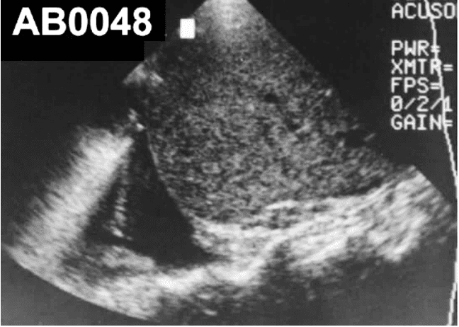
The image indicates free fluid in the pleural cavity on the right side with spine sign positive.
Explanation
Free fluid only on the right, seen above the diaphragm with a positive spine sign, indicates a right pleural effusion (in this case highly likely to be hemothorax due to trauma) and counts as a positive eFAST, even if the abdominal views are otherwise negative. In normal aerated lung, air blocks sound, so the thoracic spine is only visible below the diaphragm and “disappears” above it. When there is pleural fluid, ultrasound travels through that fluid and the vertebral bodies become visible above the diaphragm: this is the positive spine (thoracic spine) sign.
History of trauma is important to make a probable diagnosis. If there was no history of trauma then it would simple be a case of a pleural effusion. Always correlate clinically. The true confirmation would be by a CT scan if needed and checking the Hounsfield unit value of the fluid – typically in the range of roughly 30-70 HU. Other method would be to drain a sample of the fluid under ultrasound guidance.
References
Access the full POCUS Learning Library for FREE!
Share a few details so we can tailor new content to your specialty and region.


