
A 65-year-old male presented with right upper quadrant pain for few days. The following image was obtained in the right upper quadrant. Multiple gallstones were seen within the lumen of the gallbladder. On changing patient position to the left lateral and upright position the gallstones were observed to be rolling to the most dependent position within the lumen of the gallbladder. No gallstone was seen impacted in the neck of the gallbladder. Sonographic Murphy sign was negative.
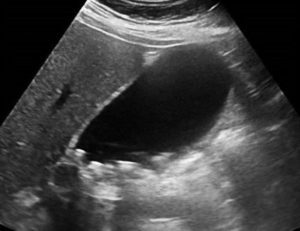
Image courtesy of UltrasoundCases.info owned by SonoSkills
What is the diagnosis?
A. Calculous cholecystitis
B. Gangrenous cholecystitis
C. Multiple gallstones/cholelithiasis
Test your knowledge of POCUS of Hepatobiliary with this knowledge check!
Answer:
Ultrasound image shows a well distended gallbladder. Multiple small gallstones seen within the lumen of the gallbladder. Gallbladder wall is not thickened. No pericholecystic fluid seen. No gallstone was seen impacted in the neck of the gallbladder. Sonographic Murphy sign was negative. No evidence of membrane like structures seen within the lumen of the gallbladder.
Reference
https://insightsimaging.springeropen.com/articles/10.1186/s13244-019-0825-4


