
Case Study:
A 67-year-old male patient came to the Emergency Department with a history of loss of vision in one eye. The patient did not complain of any pain in the affected eye. There is no history of trauma or injury to the eye. He does have a history of Myopia since childhood. He is also a known hypertensive but does take antihypertensive medication regularly. An ultrasound exam was performed on the affected eye using the ophthalmology preset. The following view was obtained.
What is the diagnosis?
A. Foreign body in the eye
B. Vitreous hemorrhage
C. Displaced intraocular lens
D. Retinal detachment
(Answer below)
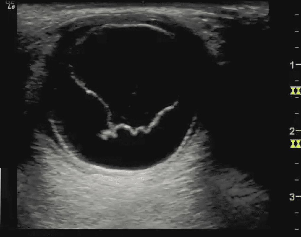
Answer: D, Retinal detachment


