
A 35-year-old female presented to the emergency department with complaint of dyspnea, discomfort while lying down, lightheadedness and slight fulness in the chest.
There was a history of mild fever for a couple of weeks. There was no history of trauma. The physician performed a limited cardiac POCUS examination. The following image was one of the images obtained.
What is the arrow pointing to?
A. Adipose tissue
B. Pleural effusion
C. Pericardial effusion
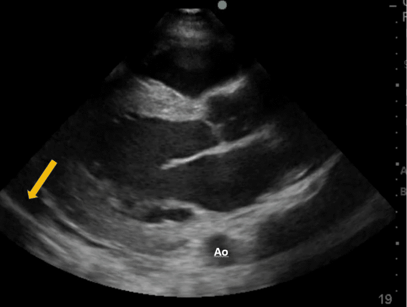
The arrow is pointing to pericardial effusion.
Explanation
The view below shows a transthoracic parasternal long axis (PLAX) view of the heart. Observe the location of the pericardium and posterior wall of the left ventricle.
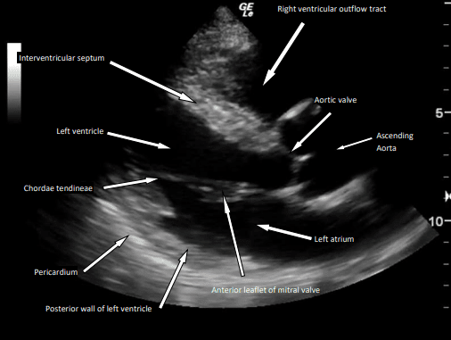
Figure 1 – PLAX view
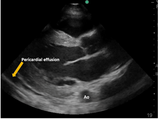
Figure 2 – The arrow is pointing to an anechoic region. This is a pericardial effusion.
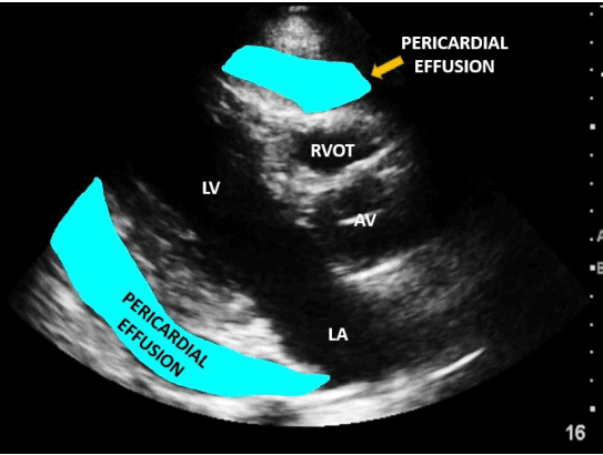
Figure 3 – pericardial effusion highlighted (another example).
References
- https://nephropocus.com/2019/12/07/pericardial-versus-pleural-effusion-on-plax-view/
- doi: 10.1016/0002-9149(80)90423-3


