A 28-year-old female was brought to the emergency department after being involved in a road traffic accident.
The patient was conscious but anxious and in pain. Heart rate was 110/minute and BP was 90/50 mm Hg. The physician on duty performed an eFAST examination. The following is one of the images obtained.
Free fluid was found in the pelvic cavity and the Morisons pouch. Considering the history, physical examination, and ultrasound findings it was determined that the patient had occult blood loss. She was immediately taken to the operating room for surgery to stop the internal bleeding.
The free fluid in the pelvic cavity is indicated by:
- A
- B
- C
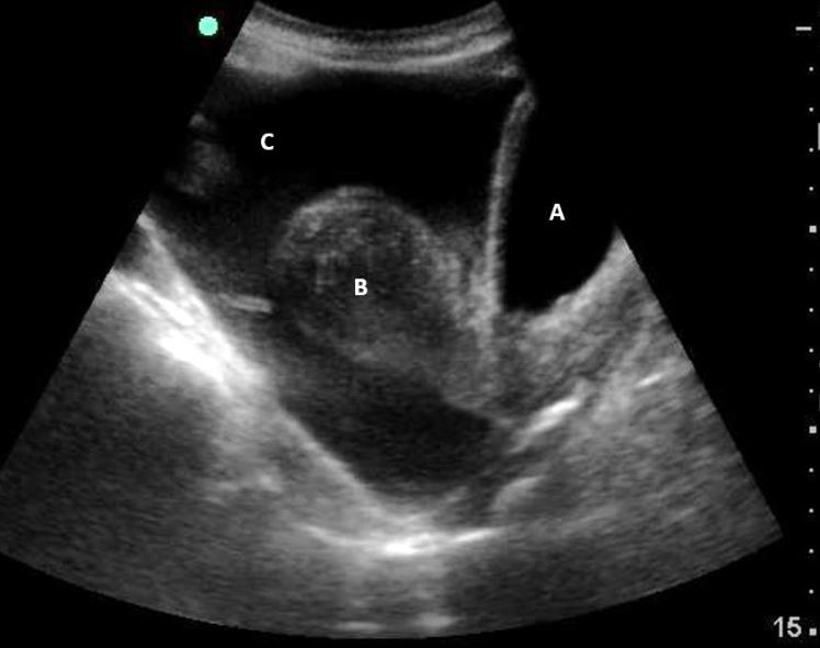
C indicates the free fluid.
Explanation
The image below shows a mid-longitudinal view of the female pelvic region. Observe the bladder (blue) partially distended with urine. The uterus is clearly seen with anechoic free fluid present anteriorly, and superiorly. Free fluid is also seen in the pouch of Douglas posterior to the uterus.
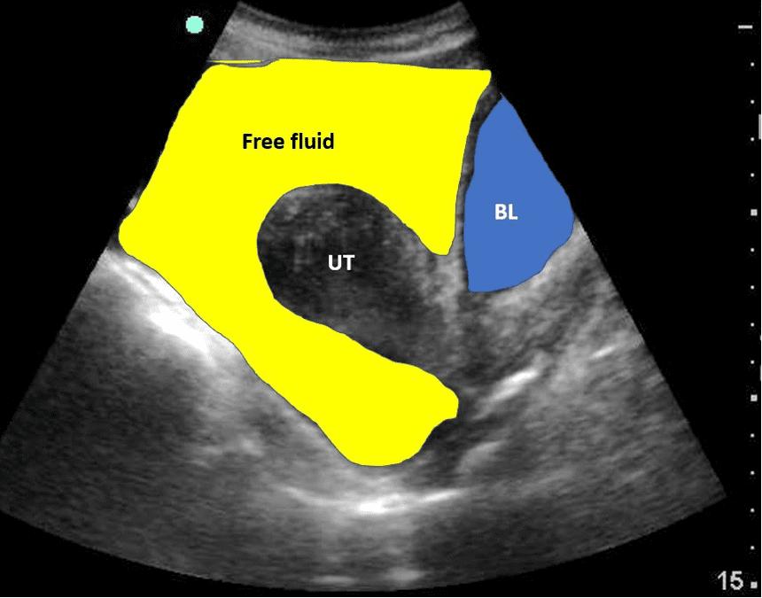
Figure 1. Mid-longitudinal view of the uterus with bladder (BL) anteriorly and inferiorly. Free fluid is highlighted in yellow. Observe the location of the uterus (UT).
References
Access the full POCUS Learning Library for FREE!
Share a few details so we can tailor new content to your specialty and region.


