
A 62-year-old male patient’s lab results showed elevated liver enzymes. There is no history of fever or abdominal pain. An ultrasound examination of the left lobe of the liver showed the view below. The caudate lobe of the liver was hypoechoic. The yellow arrow is pointing to the caudate lobe of the liver.
What is the most probable diagnosis?
A. Liver abscess
B. Liver metastasis
C. Fatty liver with focal sparing of the caudate lobe
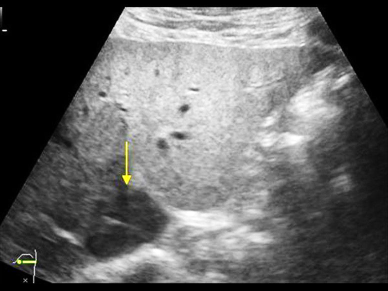
Image courtesy of UltrasoundCases.info owned by SonoSkills.
The most probable diagnosis is fatty liver with focal sparing of the caudate lobe.
Explanation
Focal areas of sparing will appear relatively hypoechoic on ultrasound as compared to the adjacent area of fatty infiltration of the liver which will appear hyperechoic. Be sure to not miss liver metastases or hepatocellular carcinoma. Always correlate clinically.
References


