
Case Study:
A 65-year-old male patient presented to the clinic for a routine physical exam. He is a known smoker. He had PMH of HTN, DM Type II and ongoing tobacco use. He did not complain of chest pain or dyspnea. The patient does complain of mild discomfort and pain in the lower abdomen, increased frequency of urination and occasional blood in the urine. He does not have any episode of passing gravel in the urine.
There is a family history of ischemic heart disease, CVA.
Temp – 98.4 degrees Fahrenheit, HR – 68 BPM, BP – 140/90 mm Hg, RR – 22 per minute
There was no other significant finding. The patient was concerned that he may have BPH. A POCUS exam was performed to examine the kidneys, ureters and bladder. The kidneys were of normal length and echotexture and showed no evidence of a mass, calculus or hydronephrosis. The bladder scan was performed as well, and the following is the longitudinal image of the bladder. The echogenic lesion inside the bladder did not move when the patient was rolled to the left and right lateral decubitus position.
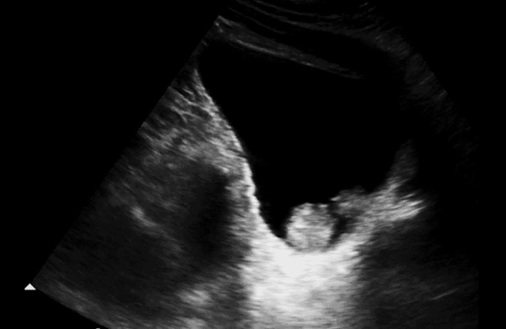
What is the most likely diagnosis?
A. BPH with enlarged median lobe protruding into the lumen of the bladder.
B. Bladder calculus
C. Normal bladder with focal contraction of detrusor muscle
D. Bladder mass – possible bladder carcinoma
Interested in proving your expertise in Genitourinary/Renal POCUS? Check out our GU/Renal POCUS Certificate here.
Answer: D, bladder mass — possible bladder carcinoma


