
An 80-year-old female presented with right upper quadrant pain and fever. She had an ERCP done the previous day. She was diagnosed with acute calculous cholecystitis and gallbladder sludge a couple of days ago and underwent ERCP. The image below was obtained during the POCUS examination.
What is the most likely diagnosis or clinical impression?
A. Emphysematous cholecystitis or air introduced into the gallbladder during ERCP
B. Gangrenous cholecystitis
C. Porcelain gallbladder
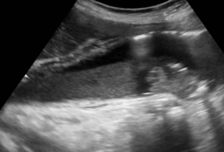 Image courtesy of UltrasoundCases.info owned by SonoSkills
The most likely diagnosis is emphysematous cholecystitis or air introduced into the gallbladder during ERCP
Explanation
The ultrasound image shows an adequately distended gallbladder with sludge layering in the dependent portion of the gallbladder. An ill-defined gallstone with posterior acoustic shadow in seen near the fundus of the gallbladder. Gallbladder wall is slightly thickened. There is a trace amount of pericholecystic fluid seen. Sonographic Murphy sign was positive. There is evidence of echogenic air within/or just under the anterior wall of the gallbladder. The air also has associated reverberation artifact and other air pocket has a dirty shadow seen posteriorly. In this case it would be difficult to comment with certainty if the air is due to emphysematous cholecystitis or due to the ERCP procedure. Perform serial follow up ultrasound exams and correlate clinically. In this case air was accidentally introduced during ERCP and papillotomy.
References
- doi: 10.1155/2017/1971457
Test your knowledge of hepatobiliary POCUS with this knowledge check!


