A 68-year-old asymptomatic male underwent abdominal aorta aneurysm (AAA) ultrasound screening.
A dilated segment of the abdominal aorta was identified in the lower abdominal aorta. The maximum outer wall to outer wall diameter of the dilated segment was 7.3 cm. The patient was immediately referred to a vascular surgeon for further investigation and management. The next day, the patient underwent endovascular aneurysm repair (EVAR). The ultrasound image below was obtained in the mid-longitudinal plane.
What type of aneurysm was identified?
A. Berry aneurysm
B. Fusiform aneurysm
C. Cannot determine from the image
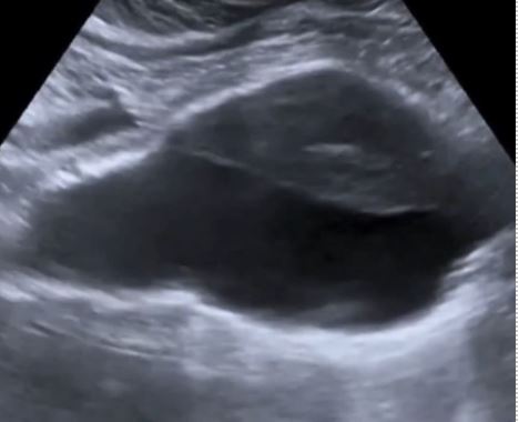
This is a fusiform aneurysm.
Explanation
The ultrasound image shows a mid-longitudinal view of the abdominal aorta through the aneurysmal segment. This is an example of a fusiform type of abdominal aortic aneurysm. See the figure below for a comparison of berry and fusiform type aneurysms.
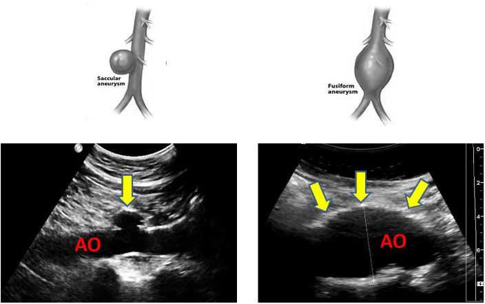
Figure 1. Types of abdominal aortic aneurysms. Saccular/berry aneurysm on the left and fusiform aneurysm on the right. Approximately 94% of aortic aneurysms are fusiform while 90% of cerebral aneurysms are saccular or berry aneurysms.
References
- https://www.uspreventiveservicestaskforce.org/uspstf/recommendation/abdominal-aortic-aneurysm-screening
- https://www.pocus101.com/aorta-ultrasound-made-easy-step-by-step-guide/
- https://sonographictendencies.com/2021/03/17/aorta-duplex-doppler-protocol/
Want to test your AAA POCUS knowledge further?
Try out our 8 question AAA knowledge check!
Access the full POCUS Learning Library for FREE!
Share a few details so we can tailor new content to your specialty and region.


