Case Study:
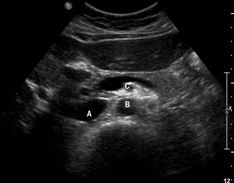
A 67-year-old male patient with a history of smoking had concerns about an abdominal aortic aneurysm (AAA). An AAA screening exam was performed. Below is the transverse view through the abdominal aorta.
Questions:
- Is there an aneurysm with an internal thrombus seen in this image?
- Was this view obtained through the upper abdominal aorta?
- Identify Vessel A, B, and C.
(Answers below)
Answers:
- No, there is no evidence of a AAA in this view. Even though the aorta was not measured. You could get an idea by comparing with the scale on the right side of the image.
- No, It is a view through the mid abdominal aorta. The SMA is visible as a circular anechoic dot surrounded by echogenic adipose tissue. So, this view was obtained below the level of the origin of the celiac axis. Thus, it was obtained at the level of the mid abdominal aorta.
- Vessel A is IVC, Vessel B is Abdominal Aorta, Vessel C is SMA
Access the full POCUS Learning Library for FREE!
Share a few details so we can tailor new content to your specialty and region.


