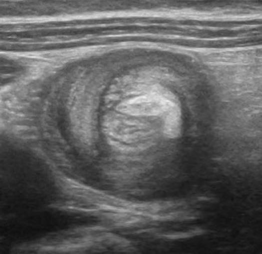
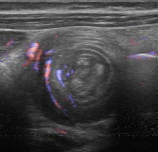
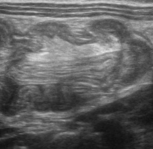
A 3-year-old female was brought to the emergency department (ED) with complaint of sudden abdominal pain, vomiting, lethargy and passage of blood and mucus in stools. A palpable lump was felt in the right lower quadrant. The mass seemed to be sausage shaped with emptiness in the right lower quadrant (Dance sign). The emergency medicine physician performed an abdominal POCUS exam. The following images were obtained in the right mid and lower abdomen.
What is the most likely diagnosis?
A. Appendicitis
B. Renal mass
C. Ileocecal intussusception


Images courtesy of UltrasoundCases.info owned by SonoSkills
The most likely diagnosis is ileocecal intussusception.
Explanation
The B-mode ultrasound image shows the doughnut sign and the crescent in doughnut sign. The hyperechoic crescent within the mass is formed by the mesentery which got dragged into the intussusception segment. The doughnut sign is seen as concentric alternating hypoechoic and hyperechoic bands. The submucosa appears hypoechoic. The hyperechoic bands represents the mucosa and muscularis layers of the bowel loops. Please see reference article for details.
References
Test your knowledge of Abdominal Trauma POCUS
with this knowledge check!


