
Can POCUS Diagnose Foreign Body or Air in Soft Tissue?
By Victor V Rao MBBS, DMRD, RDMS (APCA)
Point-of-care ultrasound (POCUS) is valuable in managing patients suspected to have foreign body lodged in their skin and soft tissue. Skin and soft tissue infections (SSTIs) can occur due to a breach of skin or its supporting structure. On a physical exam, it may be challenging to differentiate between cellulitis, abscess, folliculitis, or vesicles. The above conditions are often encountered during the physical examination. In this article we will look at some examples of foreign bodies embedded in the skin and subcutaneous tissue and look at a couple of examples of air in the subcutaneous tissue due to an infection by gas producing microorganism. Surprisingly, they may look very similar on ultrasound and therefore it is important to obtain a relevant medical history in addition to a physical examination.
The skin comprises of a superficial epidermis and a deeper, thicker layer called the dermis. The hypodermis, also known as subcutaneous fascia, is the layer deep to the dermis and is the deepest layer of skin, containing adipose tissue (fat) and subcutaneous connective tissue.
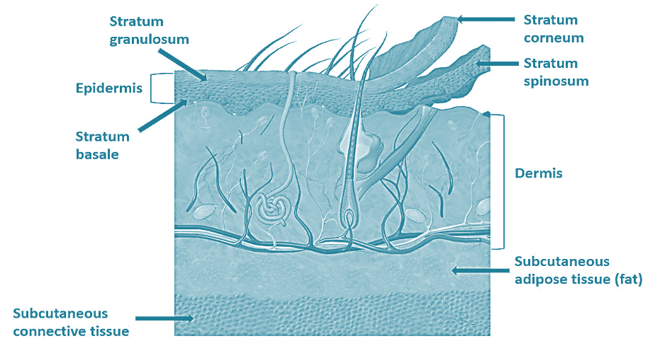
Figure: 1 Histology of skin and subcutaneous tissue
Normal Ultrasound of the Skin
Imaging the layers of the skin and tissues deeper to it is possible using a high-frequency ultrasound transducer. You may use a water bath, a thick layer of gel, or even a commercially available acoustic standoff pad during scanning to aid in improved image quality by offsetting the near zone.
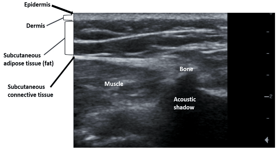
Figure 2: Normal ultrasound appearance of the skin, subcutaneous tissue, muscle, and bone. Remember that bone, foreign body, and air will appear hyperechoic on ultrasound.
Foreign Body
Medical history is very important. The patient will either provide a history of some trauma or may remember an incident where a foreign body was lodged in the skin but was removed. Sometimes, the patient may not be able to provide a clear history. Depending upon the type of foreign body, we may be able to see some echogenic structures in the layer of the skin or deeper. The following ultrasound findings may be seen:
- Increased thickness of the dermis (compared with normal side) +
- Hyperechoic structures in the skin layers or deeper.
- Evidence of edema/anechoic branching pattern (referred to as cobblestone pattern).
- Maintenance of tissue architecture (if the architecture of the soft tissue is disrupted then it is suggestive of an abscess).
- If the suspected foreign body was lodged a while ago then there may be evidence of granulation tissue around it. See Figure 4.
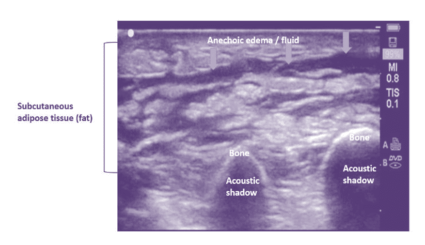
Figure 3: B-mode image shows cobblestone pattern. Subcutaneous adipose tissue appears hyperechoic with edema between fat lobules. The tissue architecture is maintained. No evidence of abscess is seen. Findings are consistent with cellulitis. Observe the normal echogenic appearance of bone with an acoustic shadow posteriorly. Do not confuse it with foreign body. Always compare with normal side.
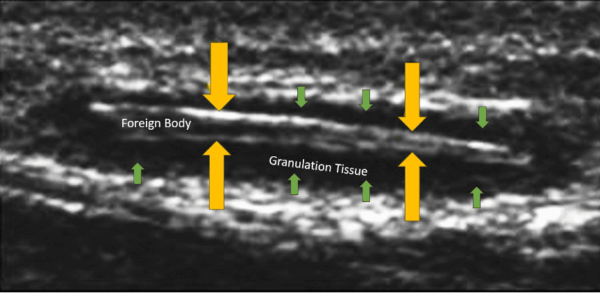
Figure 4: Linear echogenic foreign body seen with hypoechoic granulation tissue around it. If the imaging was performed within a few hours of the incident then it would be suggestive of a hematoma surrounding the foreign body. It can get tricky sometimes. Use color or power Doppler to demonstrate increased flow in the suspected region. Labeled image created using ultrasound Image courtesy of UltrasoundCases.info, owned by SonoSkills.
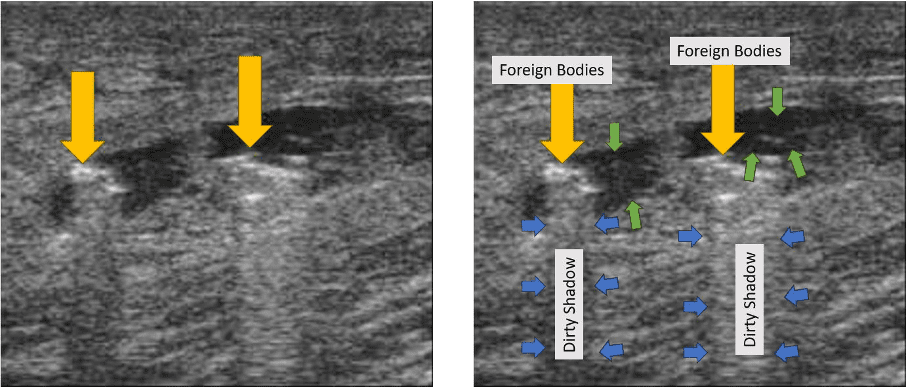
Figure 5: Multiple echogenic foreign body seen with anechoic/hypoechoic around it. This patient had multiple glass pieces embedded in the soft tissue. Since history was acute, the anechoic/hypoechoic regions surrounding or adjacent to the foreign bodies are suggestive of hematoma and not granulation tissue as seen in Fig. 4. Observe the dirty shadow like appearance posterior to the glass pieces. Labeled image created using ultrasound Image courtesy of UltrasoundCases.info, owned by SonoSkills
Soft Tissue Abscess/ Abscess/ Necrotizing Fasciitis
The following ultrasound findings may be observed in a subcutaneous abscess or a local abscess:
- Irregular or ill-defined collection.
- Disruption of normal architecture and appearance in the region with the abscess.
- Evidence of echogenic debris, septation, or even gas bubbles.
- A surrounding cobblestone pattern may still be seen if the surrounding tissue has evidence of cellulitis.
- Collection may be anechoic, hypoechoic, or heterogeneous.
- Compression with transducer may elicit movement of pus through the abscess.
- Increased surrounding vascularity is seen on color Doppler or power Doppler.
- Too much gas in the abscess may make it challenging to scan.
- Air in the soft tissue may suggest necrotizing fasciitis which is a serious condition and if not managed effectively can lead to loss of limb or even death.
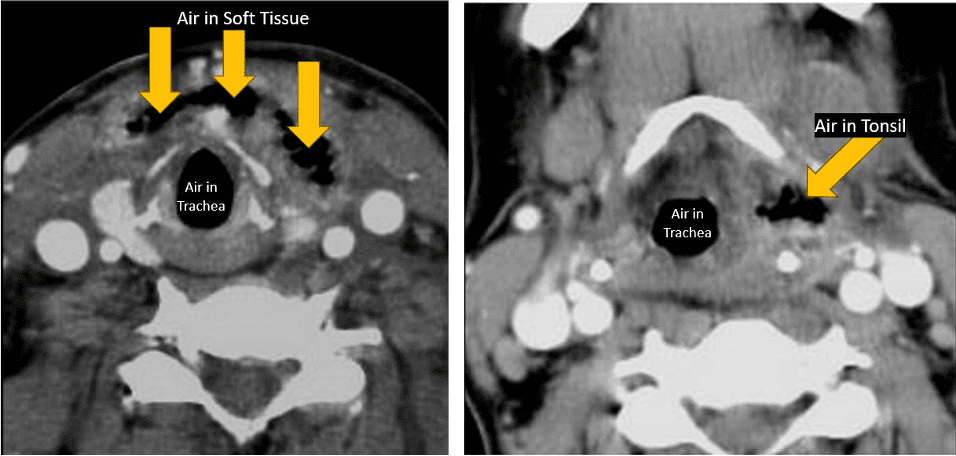
Figure 6: Axial CT with IV contrast showing presence of air (hypodense) areas in the soft tissue of the neck anterior to the trachea and in the left tonsil. These are air pockets due to gas forming organisms. See corresponding ultrasound image below. Labeled image created using ultrasound Image courtesy of UltrasoundCases.info, owned by SonoSkills.
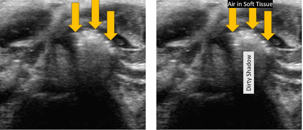
Figure 7. B-mode ultrasound of the soft tissue of the anterior neck region shows evidence of echogenic foci with dirty posterior acoustic shadows. This is due to the presence of air tracking into the tissue due to an infection by gas forming organisms. Labeled image created using ultrasound Image courtesy of UltrasoundCases.info, owned by SonoSkills.
Tips for Successful Scanning
- Select the highest possible frequency and use soft tissue or superficial preset for skin and soft tissue ultrasound scans.
- If an area of interest is very superficial consider using a thick layer of ultrasound coupling gel or an acoustic standoff or even a water bath. Make sure the transducer is not cracked and is electrically insulated before using a water bath to avoid an electrical shock.
- If you see echogenic foci in the soft tissue, make sure they are not due to air in the soft tissue which may be suggestive of necrotizing fasciitis.
- Scan in lower room lighting conditions for optimal results.
- Adjust the depth and gain settings for best results.
- Always compare with the normal side.
- Use color or power Doppler when indicated.
In conclusion, POCUS is an effective clinical tool to identify any foreign body that may be lodged in soft tissue. Use a high-frequency (approximately 10-15 MHz or higher) linear transducer and examine with both B-mode and color Doppler or power Doppler. Be vigilant to detect even tiny amount of air that may be present in soft tissue as that suggests infection by gas producing organisms and must be managed appropriately. Be aware that foreign body penetration into the soft tissue may also introduce some air into the tissue. Always correlate your ultrasound findings clinically and whenever possible do obtain a relevant medical history.


