
By Victor Rao MBBS, DMRD, RDMS (APCA)
The urinary bladder is a hollow distensible organ. Its primary function is to temporarily hold urine produced in the kidneys as it flows down the ureters. Approximate bladder volume is 300 – 500 cc in an average adult. The bladder has two ureteric orifices that bring the urine produced in the kidneys to the bladder via the ureters. The third opening is the urethral orifice from which the urine exits the bladder via the urethra during micturition.

Some common indications for urinary bladder point-of-care ultrasound (POCUS) examination are:
- Bladder volume estimation
- Bladder mass
- Bladder outlet obstruction
- Hematuria
- Hydronephrosis
- Anuria
- Flank or pelvic pain
- Confirm proper placement of Foley catheter
The low frequency curvilinear transducer is ideal for scanning the bladder (BL). If the curvilinear transducer is not available, you may also consider the phased array low frequency transducer.
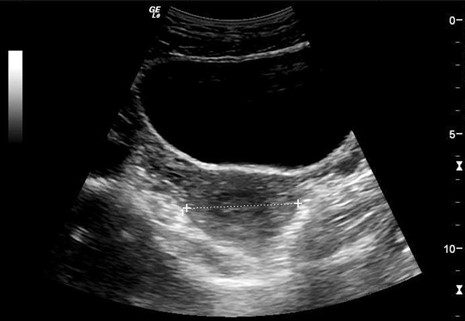
Figure 1. Transverse view of the urinary bladder. The uterus is seen posteriorly.
A full bladder also provides an excellent acoustic window to view the uterus and other pelvic organs, as seen in figure 1. Scan with the patient in the supine position. While obtaining a mid-longitudinal view of the bladder, scan with the transducer orientation marker pointing towards the patient’s head. While scanning in the transverse plane, point the transducer orientation marker towards the patient’s right side.
Bladder volume is commonly estimated while scanning the bladder. It is easy to calculate using the method shown in the figure below. Measure the anteroposterior (AP), transverse (TR), and height of the bladder in centimeters and multiply all three dimensions and then multiply with a correction factor of 0.532 to determine the volume of urine in cubic centimeters (cc). Some ultrasound devices offer automated volume estimation as well.
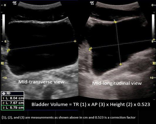
Figure 2. Bladder volume calculation using the ellipse method.
Bladder mass is another common finding that may be encountered, with transitional cell carcinoma being the most typical form of bladder cancer. Some patients are asymptomatic. See figure below.
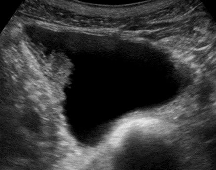
Figure 3. Bladder mass. Image courtesy of UltrasoundCases.info owned by SonoSkills.
A bladder diverticulum is generally an incidental finding on ultrasound. Rarely, calculus may form within the diverticulum. Sometimes, the neck of the diverticulum may also be seen as in the figure below.
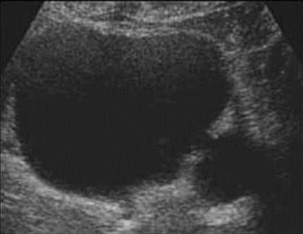
Figure 4. A small bladder diverticulum is seen arising from the defect in the bladder wall—image courtesy of UltrasoundCases.info owned by SonoSkills.
Bladder stones may be seen occasionally. You can also document the mobility of the bladder stone by rolling the patient to the left or right lateral position. The bladder stones shift position unless they are impacted at the ureterovesical junction (see figure below).
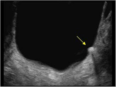
Figure 5. Single bladder calculus. Image courtesy of UltrasoundCases.info owned by SonoSkills.
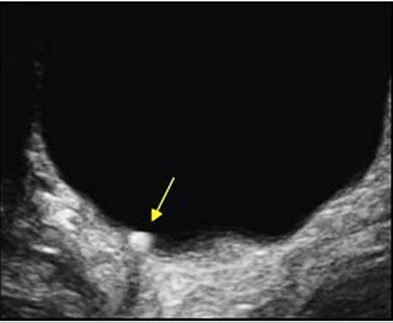
Figure 6. Bladder calculus shifted position when the patient was rolled onto the right side—image courtesy of UltrasoundCases.info owned by SonoSkills.
In conclusion, bladder ultrasound is easy to perform. Keep in mind that before conducting a scan, always obtain a brief clinical history.
Here are a few conditions where its application is most helpful. Determining the proper placement of the Foley catheter and confirming the location of the bulb of the catheter is very helpful. You can also look for ureteric jets within the bladder using color or power Doppler. Ureteric jets are important to determine if the urinary obstruction in the ureter is partial or total. If there is total obstruction, then a procedure must be performed to prevent damage to the ipsilateral kidney. In elderly males, you could also incidentally diagnose benign prostatic hyperplasia (BPH) with bladder outflow obstruction. If diagnosed early, kidney damage may be avoided, which is one of the objectives of point-of-care bladder ultrasound.
Validate your advanced knowledge of renal POCUS with the POCUS Renal/Genitourinary Certificate.
References
AIUM Practice Parameter for the Performance of an Ultrasound Examination in the Practice of Urology. (2011). The American Institute of Ultrasound in Medicine. https://www.aium.org/resources/guidelines/urology.pdf
Southgate, S. & Herbst, M. (2001). Ultrasound of the Urinary Tract. StatPearls. https://www.ncbi.nlm.nih.gov/books/NBK535381/


