
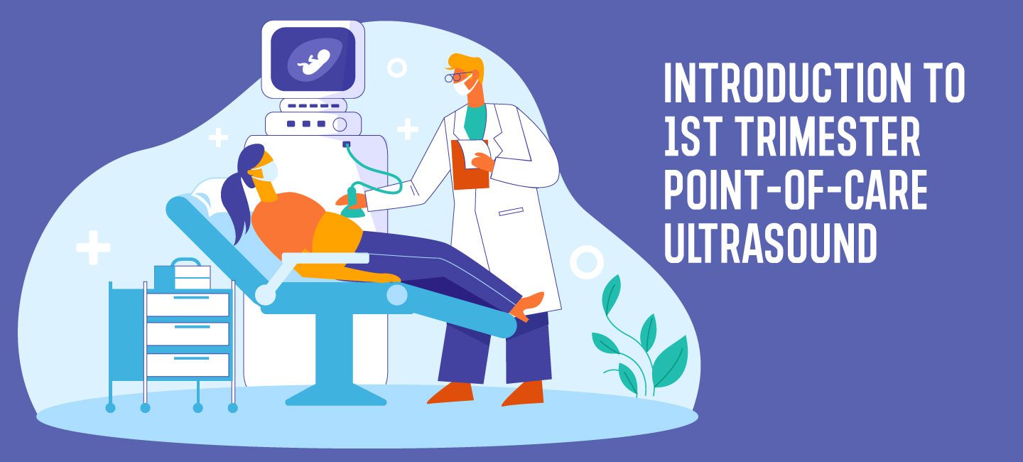
By Victor Rao MBBS, DMRD, RDMS (APCA)
The word trimester is derived from the Latin word trimestris, which is composed of two words, tri meaning “three” and mestris meaning “month.” The first-trimester point-of-care ultrasound (POCUS) examination is defined as an ultrasound examination performed up to 13 weeks gestation following the last menstrual period. During early pregnancy, which is considered the first three weeks, nothing significant can be observed during an ultrasound examination, as findings may even suggest no evidence of pregnancy.
In some patients, we are unable to visualize evidence of intrauterine pregnancy (IUP) even up to five weeks gestation using a transabdominal approach, which would require a transvaginal ultrasound examination. For this reason, a routine first-trimester ultrasound is generally performed at or after seven or eight weeks of gestation.
While performing a transabdominal scan, a full bladder is recommended. A full bladder pushes the bowel loops superiorly and provides an excellent acoustic window to image the uterus and pelvic organs. However, the bladder should be empty for a transvaginal ultrasound. During this scan, the first definitive sign of an IUP is the double decidual sac seen at five weeks gestation. The yolk sac appears next at five and half weeks and is a conclusive sign of an IUP, as seen in the image below.

Image – Courtesy: Journal of Obstetrics and Gynecology Research, Volume: 46, Issue: 2, Pages: 223-228, 2019, DOI: (10.1111/jog.14173)
During the first trimester, a POCUS examination can be performed to confirm, document, rule out or evaluate one or more of the following:
- Evidence of intrauterine pregnancy
- Ectopic or heterotopic pregnancy
- Cardiac activity
- Number of embryos
- Gestational age
- Early pregnancy failure evaluation
- Gross anomalies
- Molar pregnancy
- Determine chorionicity and number of amniotic sacs in case of multiple pregnancies
- Threatened or complete spontaneous abortion
- Patients presenting with pelvic pain and vaginal bleeding
- Pelvic mass with early pregnancy
Some clinicians believe an early first-trimester scan is not medically necessary in asymptomatic patients. However, it may be beneficial to conduct one to document the gestational age (GA), which is determined by the crown-rump length (CRL).It is important to note that CRL is the most accurate parameter used to estimate gestational age. Additional methods include using biparietal diameter (BPD), head circumference (HC), femur length (FL), or abdominal circumference (AC).
Gross fetal anomalies should be ruled out when possible. Plan a routine second-trimester scan for a more detailed look. In the case of twins or multiple pregnancies, efforts should be made to document chorionicity and amnionicity, in addition to the number of embryos.
Ectopic and heterotopic pregnancy should also be ruled out. If a patient presents with bleeding, it is important to look for the site of hemorrhage. Pregnancy viability should be documented, including cardiac activity. Always correlate ultrasound findings with ß-hCG levels.
Even though it is too early to determine if there is placenta previa or low-lying placenta, the placenta is visible.If the image shows the placenta located near and overlying the internal os, make note that follow-up scans are required.
During the first trimester, organogenesis is occurring, and it is best practice to use the ALARA principle while scanning. Do not use Doppler as this ultrasound mode has a higher acoustic output, which can heat the embryo and cause possible thermal side effects on developing embryonic tissue.
In conclusion, a first-trimester POCUS is recommended for POCUS users as it will yield valuable information. When doing so, be sure to schedule a routine second-trimester exam at 19-20 weeks gestation.
Get Started on Your Obstetrics/First Trimester POCUS Certificate Today!
References
Rodgers, S.K., Chang, C., DeBardeleben, J.T., Horrow, M.M. (2015). Normal and Abnormal US Findings in Early First-Trimester Pregnancy: Review of the Society of Radiologists in Ultrasound. RadioGraphics, volume 35 (issue 7). https://doi.org/10.1148/rg.2015150092
Sheiner, E., Freeman, J., & Abramowicz, J.S. (2005). Acoustic Output as Measured by Mechanical and Thermal Indices During Routine Obstetric Ultrasound Examinations. Journal of Ultrasound in Medicine, volume 24 (issue 12), 1665-1670. https://doi.org/10.7863/jum.2005.24.12.1665


