By Carissa Tomer, RDMS, RVT
Introduction
The uncertainty surrounding a mass in the breast often triggers significant anxiety for everyone involved including the patient. The fear of breast cancer, coupled with possible long wait times for an appointment, can make the experience even more agonizing. The benefits of timely breast mass evaluation extend beyond physical health; they can profoundly impact a patient’s mental and emotional well-being.
How does point-of-care ultrasound (POCUS) play a role in evaluating breast mass? POCUS is transforming how providers interact with and triage patients across all specialties. With a quick bedside scan, providers can gather valuable insights into the mass’s characteristics to determine whether it appears benign or suspicious by ultrasound imaging criteria.
By offering a bedside scan, healthcare practitioners can help alleviate much of the fear and anxiety that accompanies the uncertainty of a breast mass. This approach strengthens the trust between patient and provider and accelerates further evaluation when clinically necessary.
The Role of POCUS in Breast Mass Evaluation
While no ultrasound image can definitively diagnose malignancy, POCUS is a valuable triage tool to help prioritize further testing. By performing a bedside ultrasound scan, providers can offer preliminary insights into the breast mass’s ultrasound characteristics, determining whether it is potentially benign or suspicious based on several sonographic findings.
Consider a scenario where two patients present to their provider on the same day.
- Patient A: A quick scan reveals clear margins, anechoic (black) echotexture, and no suspicious features. Highly suggestive of a benign finding. A full work-up will still be ordered as a precaution; however, the patient is provided with preliminary information indicating that the breast mass observed on the initial scan appears benign.
- Patient B: The findings reveal irregular margins, posterior shadowing, and a complex echotexture. These abnormalities raise significant suspicion.
To ensure timely intervention, the provider can expedite the referral and prepare the patient mentally for a more comprehensive workup.
Furthermore, many POCUS systems offer image transmission capabilities, allowing the clinician to collaborate with a breast specialist and share ultrasound images to confirm the need for expediting care.
This approach ensures timely diagnoses for women with suspicious findings and reassures those with benign-appearing characteristics, even if they must wait for further appointments. It creates a more efficient, patient-centered process that minimizes unnecessary anxiety by addressing concerns on the spot. In the study by Balmuth et al. (2024), point-of-care ultrasound has been shown to improve patient satisfaction and provide socioemotional benefits.
Today, we can perform point-of-care ultrasound examinations like never before, and we should embrace the technology to benefit every patient we encounter. If you have the choice to go to a provider who performs POCUS and one who does not, who will you choose?
Mental Health Considerations: A Critical Perspective
From a mental health standpoint, the benefits of timely breast mass evaluation extend far beyond physical health; they can have a profound impact on a patient’s emotional well-being. As Ashley Matthews, a licensed mental health provider, has observed firsthand, the uncertainty surrounding a breast mass can exacerbate emotional distress.
Ashley shares, “I recall one patient who, after discovering a palpable mass, experienced a significant relapse of PTSD symptoms. She was unable to drive, and during the weeks she had to wait for an appointment, she required multiple emergency visits to address escalating anxiety. The prolonged uncertainty during this period led to severe emotional distress, with the patient suffering from panic attacks and debilitating fear of a diagnosis of breast cancer.”
Ashley further emphasizes, “POCUS can alleviate much of this anxiety by providing immediate insights into the mass’s nature, thereby reducing the waiting time that often contributes to mental health crises. In my experience, when patients have quick access to an ultrasound and a preliminary diagnosis, it significantly lowers their anxiety levels and helps prevent further psychological deterioration. The ability to prioritize these patients ensures timely care, benefiting their physical and emotional wellbeing”.
Understanding Ultrasound Findings
Several characteristics can help differentiate benign from suspicious findings when evaluating breast mass using ultrasound.
Here’s a quick breakdown comparing some of these ultrasound findings:

Comparing Breast Ultrasound Findings
| Benign Characteristics |
Suspicious Characteristics |
|
| Margin | Smooth, well-circumscribed | Indistinct, angular, microlobulated, or spiculated |
| Echotexture | Anechoic or homogenous | Heterogenous or complex |
| Posterior Features | Most simple cysts produce posterior enhancement | Most common: posterior shadow *Not all cancers produce a posterior shadow |
| Shape | Oval or round | Most common: irregular Less common: Oval or round |
| Orientation | Horizontal, typically wider than tall | Vertical, typically taller than wide |
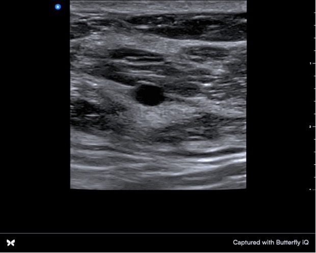
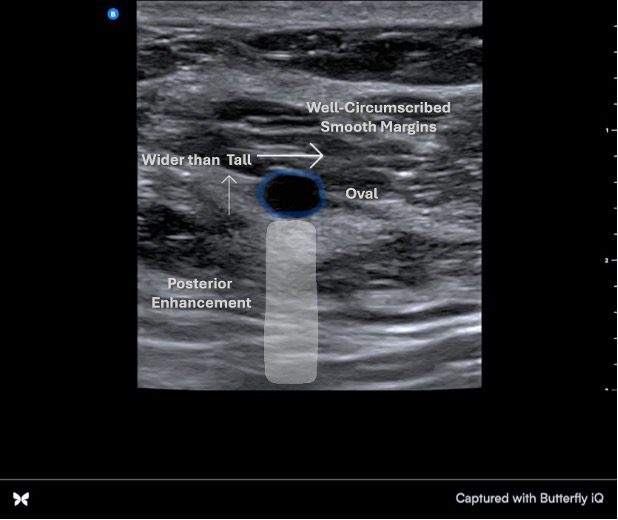
Benign Breast Ultrasound
Below is a confirmed benign, simple cyst. Note the smooth, well-circumscribed margins. Posterior image enhancement. It is oval and wider than tall. The anechoic echotexture indicates a fluid-filled center with no internal debris.
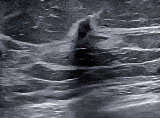
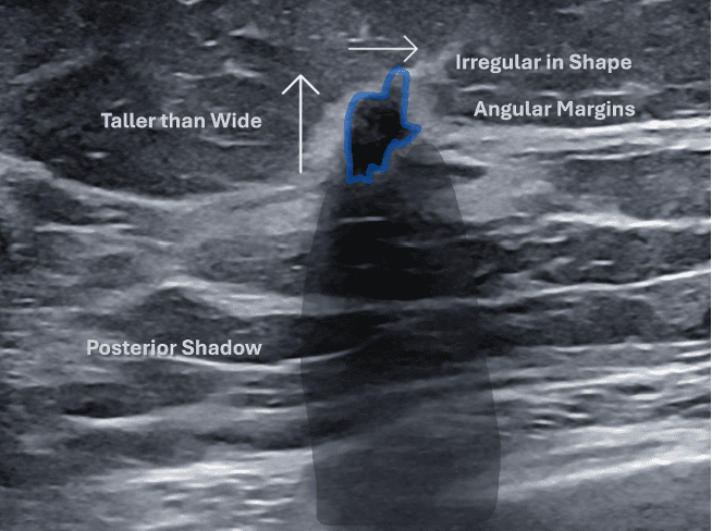
Suspicious Breast Ultrasound
Pathology confirms the lesion below as invasive ductal carcinoma. Note the angular margins and posterior shadow. It is taller than wide and irregular in shape. The internal echotexture is heterogeneous.
POCUS findings should be considered preliminary, and all palpable masses should lead to a full workup. The goal of POCUS is not to replace a full diagnostic workup but to act as an initial tool that helps prioritize patient care and reduce anxiety.
Conclusion
POCUS is an invaluable tool that provides immediate feedback. This triage approach improves the patient’s experience by alleviating anxiety and can accelerate the process of further testing. It is essential to recognize that POCUS is not a substitute for a complete workup but a triage tool that helps assess and prioritize patients. As experience with breast ultrasound grows, skills improve, enabling the delivery of more preliminary information.
Ready to learn?
Our online platform offers easy-to-follow, video-based courses designed to build your foundation. Once you’re comfortable with the basics, practice image acquisition and interpretation skills in our simulated scanning environment, where you’ll have access to thousands of pathologic cases from real patients.
What sets us apart? You’ll have a personal mentor throughout the program, available to support you and answer questions.
After completing the course, we’ll assist you in navigating the certification process, ensuring you gain the knowledge, skills, and confidence to succeed!
Let’s get you enrolled!
Visit us at UltrasoundEnergy.com
Or email us at: Clientservices@ultrasoundenergy.com
References
- Balmuth, E. A., Luan, D., Jannat-Khah, D., Evans, A., Wong, T., & Scales, D. A. (2024). Point-of-care ultrasound (POCUS): Assessing patient satisfaction and socioemotional benefits in the hospital setting. PLOS ONE, 19(2), e0298665. https://doi.org/10.1371/journal.pone.0298665


