A 45-year-old female presented to the clinic with pain on the left side and back (below the ribs).
She had complained of similar pain in the past. A kidney, ureter, bladder Point-of-Care ultrasound was performed. The following image was obtained.
What is the most likely diagnosis?
A. Renal stones/calculi in the left kidney
B. Left kidney severe hydronephrosis and hydroureter
C. Left kidney mild hydronephrosis and hydroureter
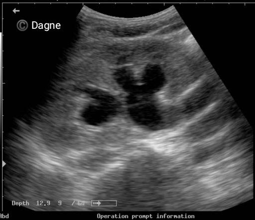
Image courtesy of Dr. Dagnechew Degefu
The most likely diagnosis is left kidney mild hydronephrosis and hydroureter.
Explanation
The image shows dilatation of the pelvicalyceal system and the upper ureter.
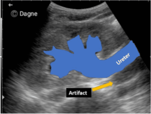
Figure 1 – Mild hydronephrosis. Observe the location of the ureter. No calculus is seen in the view. In a situation like this follow the ureter down along the mid-long axis of the ureter to locate the site of obstruction.
No calculus is seen in the image provided. There could either be an intrinsic obstruction due to a calculus or an extrinsic compression due to a mass etc. Scan should be performed along the length of the ureter to locate the site of obstruction. The calyces are dilated but still show concavity in their contour. Renal parenchymal thickness is also preserved.

Figure 2 – Normal pelvicalyceal system and different grades of hydronephrosis.
The figure shows one type of grading system. There are some other grading systems for hydronephrosis in literature. This simple straightforward grading system works well for POCUS applications.
References


