A 28-year-old female presented with nausea, vomiting and pain in the right lower abdomen. An abdominal POCUS was performed by the clinician. The following images were obtained with compression.
What is the most likely diagnosis?
A. Intussusception
B. Bowel diverticulum
C. Acute appendicitis
Acute appendicitis is the most likely diagnosis.
Explanation
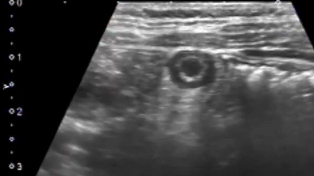
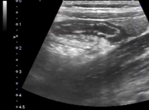
The image shows a dilated noncompressible appendix. Even though the measurement of the appendix diameter is not shown in the images, we can still determine the diameter of the appendix by using the scale seen in the image on the left side. Ideally, we should measure the maximum diameter of the dilated appendix segment from outer wall to outer wall during scanning and document it. Appendix diameter >6.0 mm is diagnostic of acute appendicitis. Target sign is positive in the transverse view. No evidence of a complex collection is seen around the appendix suggesting an intact appendix (not perforated yet). No appendicolith is seen.
References
Test your knowledge of Abdominal Trauma POCUS
with this knowledge check!


