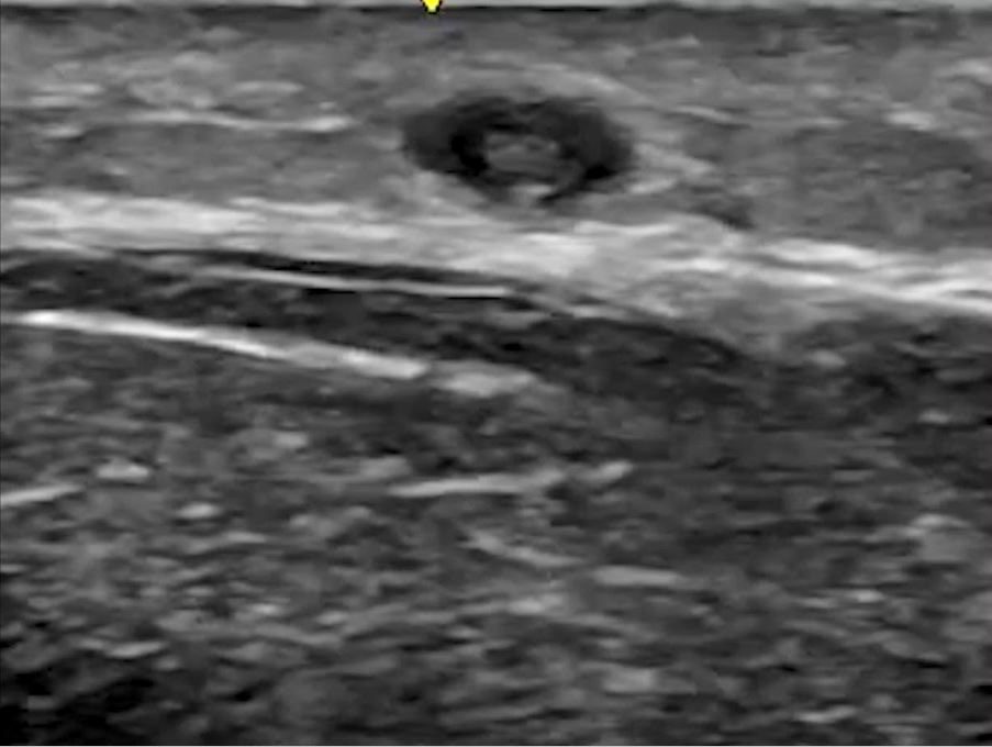
A 23-year-old male patient was admitted in the hospital and was receiving IV fluids and IV antibiotics. He complained of pain in the forearm where the IV was being administered. A POCUS soft tissue ultrasound was performed. The following image and video were obtained at the site of pain.
What is the most likely diagnosis?
A. DVT
B. Hematoma
C. Superficial vein thrombosis
Explanation
Superficial veins are not accompanied by a corresponding artery. In the video and image, we only see a single vessel. The video shows a superficial vein with some echogenicity within it suggestive of a blood clot. The compression test is more reliable and shows a non-compressible superficial vein.
See the video below of a normal fully compressible superficial vein. We generally do not use ultrasound to diagnose superficial vein thrombosis. If you are planning to insert an IV cannula and have doubt about the patency of a superficial vein, you may consider scanning the superficial vein with a high frequency linear ultrasound transducer and performing a compression test. The normal vein should collapse completely with compression force applied with the transducer.
Reference
https://radiopaedia.org/articles/superficial-thrombophlebitis?lang=us
Want to test your MSK POCUS knowledge further?
Try out our 7 question MSK knowledge check!


