A 45-year-old male presented to the clinic with complaint of pain, swelling, redness on the dorsum of the right foot for 5 days. The following ultrasound image was obtained while performing a soft tissue MSK ultrasound examination using a high frequency linear transducer and the soft tissue MSK preset.
What is the most likely diagnosis?
A. Hematoma
B. Cellulitis
C. Abscess
Answer
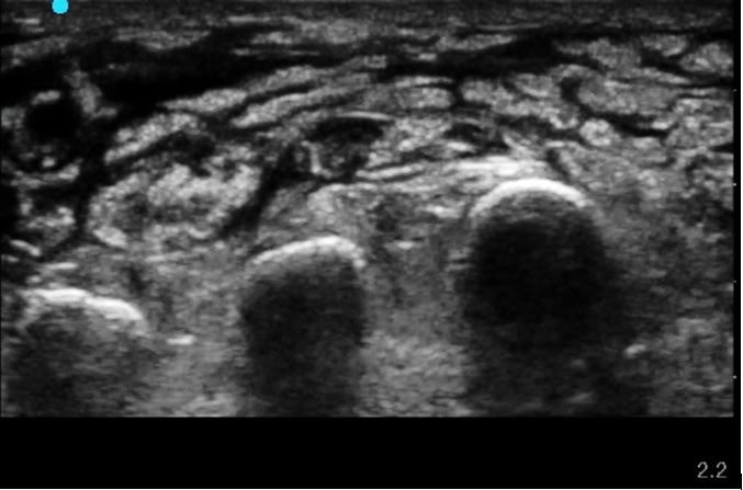
Soft tissue B-mode ultrasound image shows evidence of soft tissue swelling, anechoic fluid with a classic cobblestone pattern. There is no evidence of an abscess. Echogenic bony landmarks seen with dense acoustic shadow in the far field.
References:


