Victor V Rao MBBS, DMRD, RDMS (APCA)
Kidney stones, or renal calculi, are one of the most common health conditions patients experience in the urinary system. While approximately 90% of renal system calculi are radiopaque, 10% may not be detectable on plain abdomen X-rays or CT scans. However, an ultrasound can detect renal calculi with a specificity of 97% and sensitivity of 67%. Situations like these make point-of-care ultrasound (POCUS) an incredibly useful tool in assessing kidney, ureter, and urinary bladder conditions. By allowing clinicians to immediately perform an exam and view imaging at the patients’ bedside, POCUS may help speed the process of locating and diagnosing calculi.
The ultrasound exam of the urinary system is relatively simple to perform. Instruct the patient to fast overnight before the exam and ensure they have a full bladder during the exam. Immediate post-void bladder volume can then also be estimated.
When assessing the urinary system, the ideal transducer to use is a low-frequency curvilinear transducer. The next best option is a low-frequency phased array transducer. If you use a multifrequency single transducer device, select the abdomen or renal preset to optimize the device for best results.
While the exam is simple, calculi appear differently in different parts of the urinary system. Here are some things to keep in mind when attempting to diagnose calculi:
- Urinary calculus appears hyperechoic on diagnostic ultrasound imaging and may cast an acoustic shadow. These shadows often vary due to the size and composition of the stone. The calculus may show a twinkle artifact when scanned with the color Doppler.
- Calculi in the pelvicalyceal system may be asymptomatic; however, they can cause pain by obstructing urine flow into the ureters.
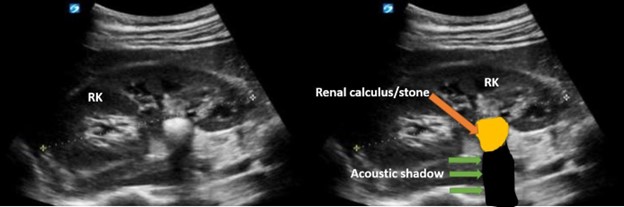
Figure 1 shows a single large hyperechoic calculus in the renal pelvis. Observe the posterior acoustic shadow due to the stone.
- On the other hand, ureteric calculi often cause severe pain described as renal colic. Patients may experience nausea, vomiting, and hematuria. The hematuria may be microscopic–invisible to the naked eye. Calculi can be seen within the ureter at any level but tend to be impacted at three potential sites of narrowing in the ureter. Those sites are located in the ureteropelvic junction (UPJ), the level where the ureter crosses anterior to the pelvic vessels, and the ureterovesical junction (UVJ).
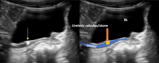
Figure 2 shows a single hyperechoic calculus in the ureter. Image courtesy of UltrasoundCases.info owned by SonoSkills.
- Calculi within the urinary bladder may be asymptomatic but often become symptomatic if the calculi are obstructed in the urethra
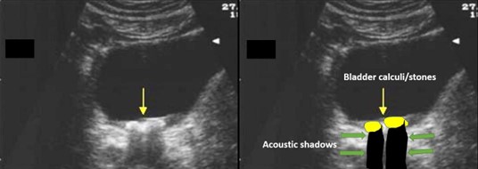
Figure 3 shows hyperechoic calculi within the lumen of the urinary bladder. You can the roll patient to the lateral decubitus position to document mobility. Image courtesy of UltrasoundCases.info owned by SonoSkills.
- If the bladder diverticulum is present, observe for the presence of calculi within the diverticulum.
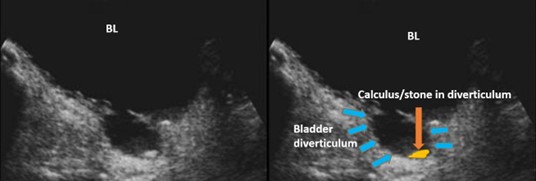
Figure 4 shows a single calculus within the lumen of the bladder diverticulum. Image courtesy of UltrasoundCases.info owned by SonoSkills.
POCUS is a great tool to diagnose the presence of stones within the urinary system. With the modality, clinicians can quickly and accurately locate the source of the problem. Pain associated with renal calculi has often been misdiagnosed as general lower back pain, but with POCUS imaging, examiners know how to help their patients accurately. Additionally, pitfalls such as bowel gas can prevent visualization of urinary calculi, but using color Doppler to recognize the twinkle artifact can easily enhance diagnostic confidence.
Thousands of people experience pain caused by calculi each year, and with POCUS, clinicians can rapidly begin effective treatment for their patients.


